Journal List > J Korean Foot Ankle Soc > v.24(4) > 1145874

만성 비골건 재발성 탈구에서 원위 비골 회전 성형술: 술기 보고

Abstract

Chronic recurrent peroneal dislocation often responds poorly to conservative treatment. Surgical treatment has been reported to be more effective than conservative treatment, and various surgical treatment methods are available: superior peroneal retinaculum repair or reattachment, peroneal groove deepening procedures, rerouting procedures, or bone block procedures. Although various treatment options have been reported, there is no consensus regarding which treatment is better. This paper proposes a distal fibular rotational plasty that can prevent recurrent peroneal dislocations and recover its function well by securing a stable peroneal tendon excursion space.

만성 비골건 재발성 탈구는 흔하지 않지만 진단이 늦어지기 쉽고,운동 능력에 상당한 제한을 주게 된다.1,2) 보존적 치료보다는 주로 수술적 치료로 좋은 결과를 얻을 수 있으며 다양한 수술방법이 보고된 바 있다.3,4) 그중 상비골건지대(superior peroneal retinaculum) 를 봉합하거나 비골구 심화술(peroneal groove deepening procedure)을 동시에 시행하는 술기가 많이 보고되었고 bone block procedure, rerouting method 등이 보고되었다.5-8) 아직 어떤 술기가 가장 우선시되는 술기인지는 이견이 있으며 상비골건지대가 확실하게 재건되고 비골건 주행 통로의 공간이 안정적이고 충분한 공간이 되어야 한다는 쪽으로 의견이 모아지고 있는 상태이다.9) 상비골건지대의 봉합 시행 시 안정적인 봉합을 위해 지나치게 단단한 봉합으로 비골건 주행 통로의 압박이 유발될 경우 통증이 유발될 수 있어 봉합 시에 주의가 요구된다.3,10) 이에 비골구 심화술 등 골성구조에 대한 수술은 비골건 주행 통로의 공간을 확보할 수 있어 봉합술과 동시에 많이 시행되고 있다.9) 하지만 비골건의 주요 탈구가 일어나는 지점이 원위비골의 후외측 첨부로 생각되는데 기존의 비골구 심화술로는 원위 비골 첨부까지 심화(deepening)가 잘 되지 않음을 저자들은 경험한 바 있다. 또한 기존의 bone block procedure 에서도 원위비골 첨부의 후외측에 지지구조가 형성되지 않거나 절골술 시 후방 비골구의 비골건 주행 표면의 침범이나 주변 인대 손상에 대한 우려가 있음을 발견하였다.9,10) 따라서 본 저자들은 골성 구조에 대한 수술을 통해 원위비골 첨부 후외측 부위에 재탈구를 막을 수 있는 안정적인 구조를 형성하면서 비골건이 안정적으로 주행하는 충분한 공간을 확보할 수 있는 원위 비골 회전 성형술을 소개하고자 한다. 본 연구는 본원의 기관생명윤리심의위원회(Institutional Review Board)의 승인을 받았다.

술 기

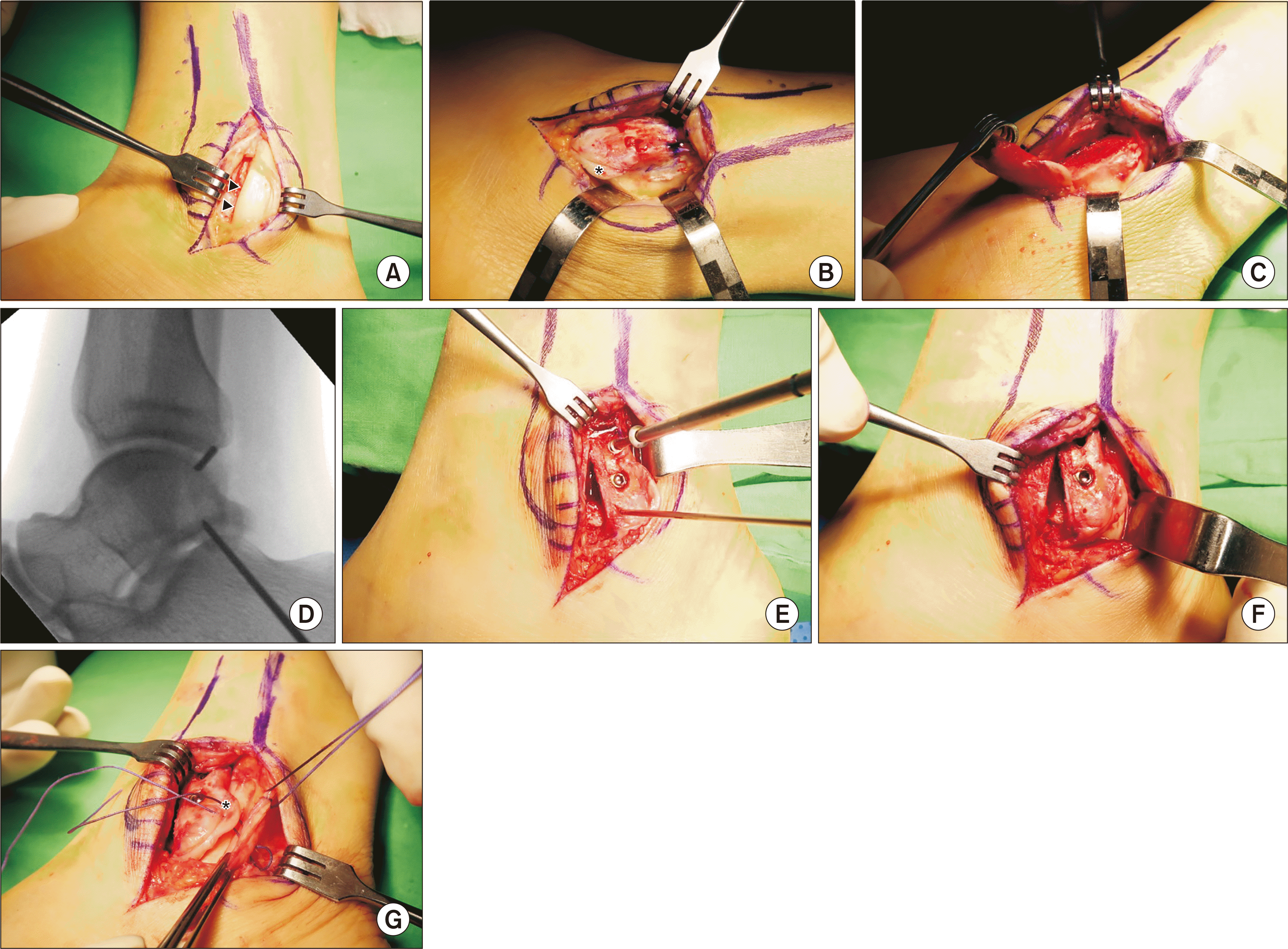
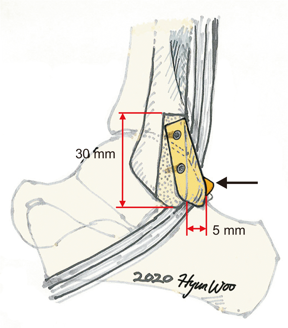
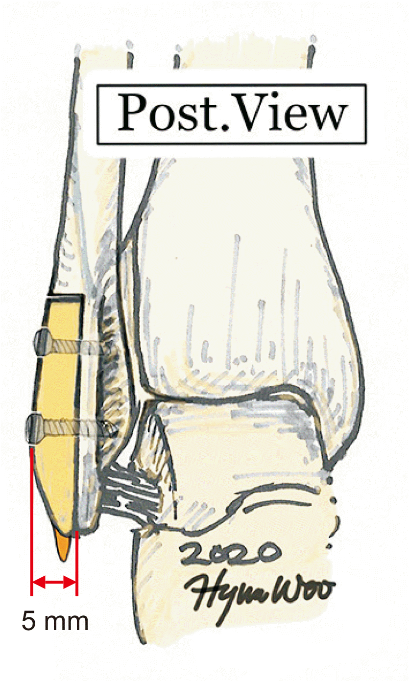
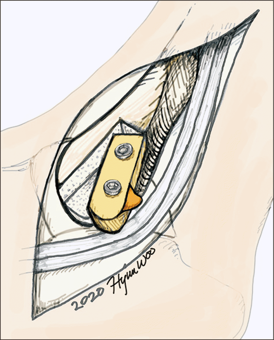
척추 또는 전신 마취를 시행한 후 측와위에서 환측 대퇴 근위부에 지혈대를 적용한 후, 원위 비골 중앙으로 후연에 가깝게 피부를 6 cm 가량 종절개한 후 원위 비골 후외측연에서 3 mm 정도의 근막 cuff를 남긴 후 근막을 절개하자 탈구된 비골건을 발견할 수 있었다 (Fig. 1A). 이때 모든 환자들에서 비골건이 탈출하면서 만든 근막과 골막 사이에 주머니 공간(pocket)이 발견되며 외과의 후연으로 섬유 연골능선(fibrocartilaginous ridge)이 관찰되며(Fig. 1B) 여기에 연결이 있던 상비골지대가 늘어나거나 연결이 단절된 소견을 보인다. 동반된 비골건의 파열이나 추가적인 소견이 있는지 확인한 뒤 건을 탈구시켜 외측으로 젖히고 난 후 원위 비골의 중앙선을 따라 원위 비골 첨부에서 근위 3 cm의 길이와 5 mm 두께로 절골을 시행하였다 (Fig. 2, 3, 4A). 또한 비골구 후방에서 섬유연골능선을 포함해 비골 구의 외측 가장자리에서 절골을 하여 최대한 비골구의 섬유연골 표면을 보존하였다(Fig. 1C). 골편을 약 15∼20도 회전시켜 5 mm 이상의 비골건의 탈구를 막는 지붕(roof)을 형성한 후 K강선으로 임시 고정하였다(Fig. 1D, 2, 4B, 5). 비골건이 족관절 신전 및 외전 시에도 탈구되지 않고 부드럽게 주행함을 확인 후 4.0 mm 해면골 나사 (cancellous screw) 두 개를 이용하여 압박 고정하였다(Fig. 1E, F).
이때 근위부의 나사는 절골면에 수직으로 삽입하였으며 원위부의 나사는 뒤쪽 외측 원위 골편에서 앞쪽 내측 원위 비골 방향으로 사선으로 삽입하였다(Fig. 1D, E). 나사의 고정이 단단함을 확인하고 C형 영상증폭기를 이용하여 나사가 관절 내로 침범하지 않음을 확인하였 다. 이후 늘어난 상비골건지대와 근막이 후방(posterior) flap이 되고, 전방(anterior)에는 섬유연골능선을 서로 봉합하여 상비골건지대를 재건하고 그 위로 절개된 근막끼리 봉합하였다(Fig. 1G). 다시 한번 비골건이 스트레스 시에도 탈구되지 않고 부드럽게 주행함을 확인한 후 피부 봉합을 시행하였다.
수술 후 4주간 단하지 석고고정 및 부분체중 부하를 유지하였으며 이후 적극적인 발목 관절 운동 및 4주간 밴드형 발목 보호대(ankle band) 착용 후 전체중 부하를 시행하였고 스포츠 활동은 수술 후 10 주까지 제한하였다.

고 찰

만성 비골건 재발성 탈구의 수술적 치료법은 다양하고 좋은 임상 결과를 보고하고 있지만 드문 질환으로 연구가 부족한 상태로 어떤 수술적 치료법이 우수한 지 비교한 연구결과가 많지 않다.5,8) 비골건이 재탈구되는 것을 막아주기 위해 상비골건지대를 안정적인 구조로 봉합하는 것이 중요하고 비골구 심화술, bone block procedure 등을 같이 시행하는 것은 비골건의 주행 시 충분한 공간 구조를 확보하고 주행 공간 내의 압력을 낮춰 재발을 막기 위한 이유이다.2,9) 본 저자들이 제안한 원위 비골 회전 성형술은 상기의 목표를 달성하면서 재탈구가 일어나는 것을 효과적으로 막기위해 원위 비골 후외측 첨부에 안정적인 골성 구조를 형성하였다.
Bone block procedure로는 원위 비골에서 15 mm 너비(width) 로 쐐기골(bone wedge)을 떼어 7 mm 후방으로 이동시켜 나사 고정하는 Du Vries procedure가 소개된 바 있으며, Tomihara 등6)은 13.3% (2/15)에서 쐐기골 혹은 나사로 인한 수술 후 통증, 6.7% (1/15)에서 수술 후 비골의 골절, 13.3% (2/15)에서 수술 후 재탈구를 경험하였다고 보고하였다.6) 하지만 Zhenbo 등9)은 원위 비골 근위 3cm부터 첨부까지 20도의 경사 절골술 시행 후 3∼5 mm 후방 으로 이동시켜 2∼3개의 흡수성 나사로 고정하는 활주 비골 이식술 (sliding fibular graft)을 26예의 증례에서 시행하였으며 수술 후 재탈구와 활주 비골 이식 골편이나 나사로 인한 수술 후 통증은 없었고 88.5%에서 excellent와 good의 만족스러운 좋은 결과를 보고하였다. 11.5% (3/26)에서 피로골절 소견이 있었으나 이들은 보존적 치료 후 호전되었다고 보고하였다.9) 하지만 원위 비골 절골술 시 전거 비인대의 손상에 주의해야할 것이며 전방에서 후방으로 절골술 시 후방 비골구의 주행표면을 침범하지 않도록 주의를 요하는 술기로 생각된다.
최근 Deng 등10)은 상비골건지대 재부착술 단독 시행군과 bone block procedure를 동시에 시행한 군의 비교 연구를 발표하였다. Bone block procedure로는 원위 비골 첨부는 보존하고 3 mm 두께 로 전방은 침범하지 않으면서 피질골(bone block) (20×15×3 mm3) 을 뗀 후 45도 후방으로 회전 후 비흡수성 봉합사로 비골 골막에 봉 합하여 고정하는 술기를 제시하였다.10) 두 군 모두 90% 이상의 만족도를 보였고 작은 피질골로 인한 자극 증상 및 통증은 관찰되지 않았으며 스트레스 골절이나 지연유합과 같은 합병증은 없었다고 하였다. 수술 후 재발에 있어서 상비골건지대 봉합군에서는 없었으며 bone block procedure군은 8.3% (2/24)였으나 통계적으로 유의한 결과는 아니었다. 결론적으로 스포츠 활동으로 복귀시점에서 상비골 건지대 봉합군이 유의하게 빨랐으며(6개월[4.3, 6.0] vs. 5개월[4.0, 5.0]), 그 외에는 두 군 간의 합병증과 임상 결과에서 차이가 없었다고 보고하였다. 전거비인대를 보존할 수 있고 피질골이 얇고 작아 이로 인한 자극 증상 및 불편감 등은 적을 것으로 보이나 피로 골절 및 골유합에 있어 좀더 연구가 필요할 것으로 생각된다.
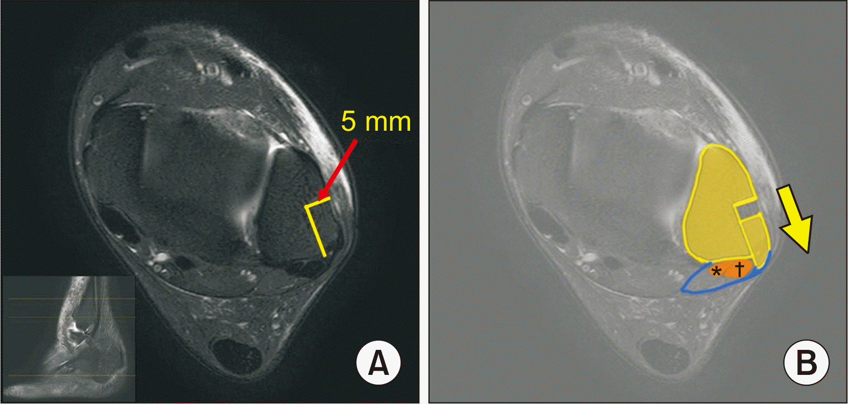
본 저자들은 Zhenbo 등9)의 술기처럼 원위 비골 첨부의 후외측 부위에 비골건의 탈구를 막을 수 있는 구조를 형성하고, Deng 등10)의 술기와 같이 주변인대의 손상 및 후방 비골구의 주행표면을 최대한 보존하는 절골술을 제시하였다. 비골건을 외측으로 젖히고 비골구 후방에서 비골건 주행 표면의 가장 외측 가장자리를 확인 후 섬유연 골능선을 포함하여 절골하여 최대한 비골구 섬유연골 표면을 보존하 였다. 골편으로 형성한 비골건 탈구를 막는 지붕은 5 mm를 넘지 않도록 하여 회전하여 고정하였으며 나사 고정 시 안정성 및 골유합을 위해 2개의 해면골 나사를 사용하였고 C형 영상 증폭기를 확인하여 나사 끝이 관절내로 침범하지 않도록 주의하였다.
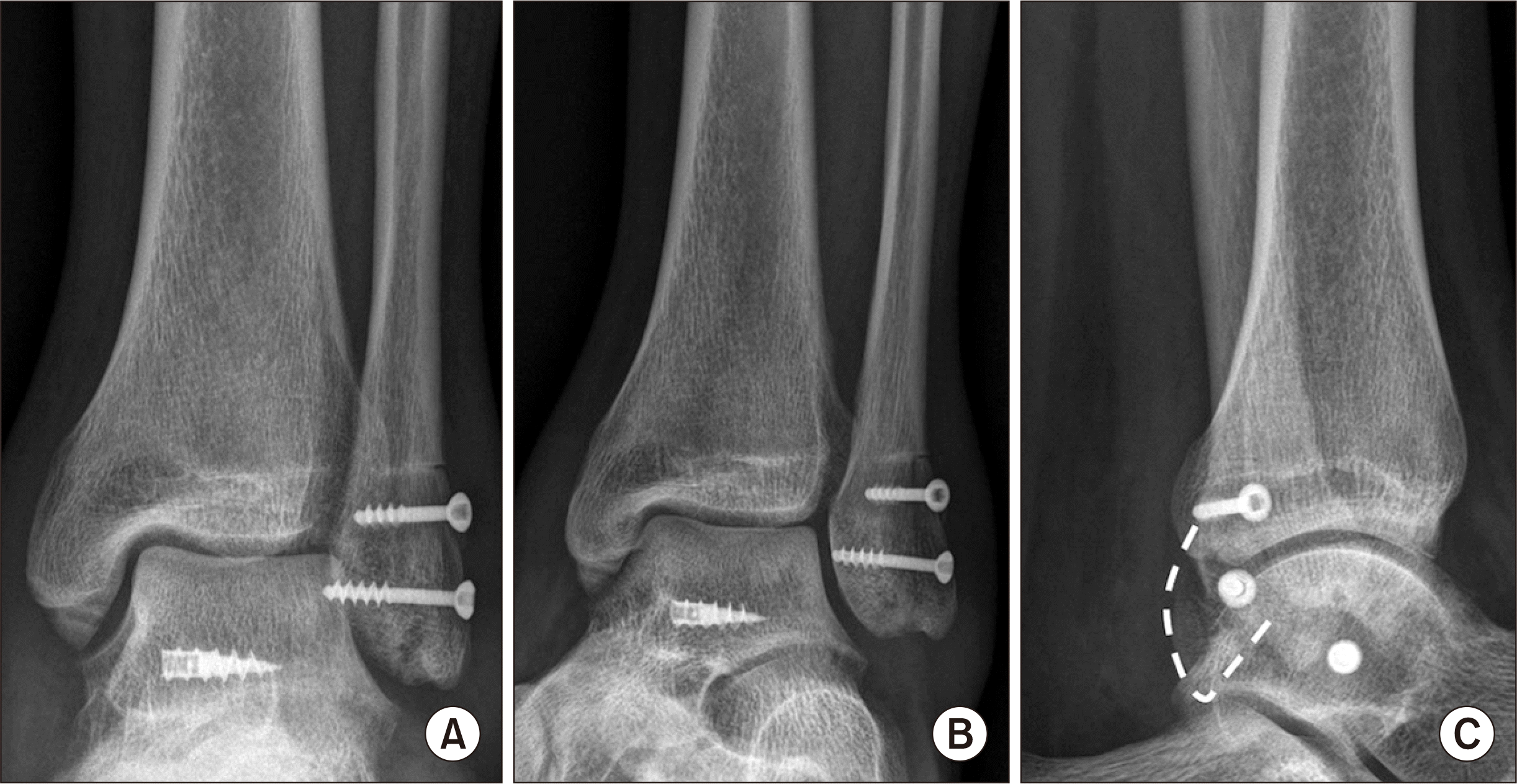
본 저자들의 경험한 증례에서 29세 남자환자는 수술 후 1년째 American Orthopaedic Foot and Ankle Society (AOFAS) 점수 87점이었으며 수술 후 재탈구 및 나사, 골편으로 인한 불편감은 없었으며, 수술 만족도상 재수술 및 타인 추천 점수가 각 10점(10점 만 점)으로 높았다(Fig. 6).
본 술식은 기존 골성 구조에 대한 수술에 비해 나사 고정이 필요하 다는 점, 그로 인해 발생할 수 있는 통증 및 자극 증상 등은 피할 수 없는 제한점이나 안정적인 유합을 이루기 해서는 필요하다고 생각하였다. Zhenbo 등9) 은 피질골 고정을 위해 흡수성 나사를 사용했었고 나사에 의한 통증은 없었으나 19.2% (5/26)에서 지연유합을 경험하였고 이로 인해 회복기간이 길어졌음을 보고하였다. 지연유합을 예방하고 일상 생활 및 스포츠 활동으로 복귀가 늦어지지 않도록 나사 고정으로 안정적인 유합을 이루는 것이 중요하다고 생각한다. 본 저자들이 시행한 증례에서는 불유합이나 지연유합은 없었으나 이에 대해서는 추가적인 연구가 필요할 것으로 생각한다.
만성 비골건 재발성 탈구의 수술적 치료에서 원위 비골의 회전 골성형술은 안정적인 비골건 주행 공간의 확보를 통해 재발을 막고 기능의 회복을 기대할 수 있는 술기로 생각한다.

Notes

Financial support: None.

Conflict of interest: None.

REFERENCES

1. van Dijk PAD, Vopat BG, Guss D, Younger A, DiGiovanni CW. 2017; Retromalleolar groove deepening in recurrent peroneal tendon dislocation: technique tip. Orthop J Sports Med. 5:2325967117706670. doi: 10.1177/2325967117706673. DOI: 10.1177/2325967117706673. PMID: 28540318. PMCID: PMC5431455.
crossref
2. Suh JW, Lee JW, Park JY, Choi WJ, Han SH. 2018; Posterior fibular groove deepening procedure with low-profile screw fixation of fibrocartilaginous flap for chronic peroneal tendon dislocation. J Foot Ankle Surg. 57:478–83. doi: 10.1053/j.jfas.2017.10.033. DOI: 10.1053/j.jfas.2017.10.033. PMID: 29269024.
crossref
3. Cho J, Kim JY, Song DG, Lee WC. 2014; Comparison of outcome after retinaculum repair with and without fibular groove deepening for recurrent dislocation of the peroneal tendons. Foot Ankle Int. 35:683–9. doi: 10.1177/1071100714531233. DOI: 10.1177/1071100714531233. PMID: 24709746.
crossref
4. Heckman DS, Reddy S, Pedowitz D, Wapner KL, Parekh SG. 2008; Operative treatment for peroneal tendon disorders. J Bone Joint Surg Am. 90:404–18. doi: 10.2106/JBJS.G.00965. DOI: 10.2106/JBJS.G.00965. PMID: 18245603.
crossref
5. van Dijk PA, Gianakos AL, Kerkhoffs GM, Kennedy JG. 2016; Return to sports and clinical outcomes in patients treated for peroneal tendon dislocation: a systematic review. Knee Surg Sports Traumatol Arthrosc. 24:1155–64. doi: 10.1007/s00167-015-3833-z. DOI: 10.1007/s00167-015-3833-z. PMID: 26519186. PMCID: PMC4823328.
crossref
6. Tomihara T, Shimada N, Yoshida G, Kaneda K, Matsuura T, Satake S. 2010; Comparison of modified Das De procedure with Du Vries procedure for traumatic peroneal tendon dislocation. Arch Orthop Trauma Surg. 130:1059–63. doi: 10.1007/s00402-010-1136-x. DOI: 10.1007/s00402-010-1136-x. PMID: 20556617.
crossref
7. Wang CC, Wang SJ, Lien SB, Lin LC. 2009; A new peroneal tendon rerouting method to treat recurrent dislocation of peroneal tendons. Am J Sports Med. 37:552–7. doi: 10.1177/0363546508325924. DOI: 10.1177/0363546508325924. PMID: 19059898.
crossref
8. Ogawa BK, Thordarson DB. 2007; Current concepts review: peroneal tendon subluxation and dislocation. Foot Ankle Int. 28:1034–40. doi: 10.3113/FAI.2007.1034. DOI: 10.3113/FAI.2007.1034. PMID: 17880883.
9. Zhenbo Z, Jin W, Haifeng G, Huanting L, Feng C, Ming L. 2014; Sliding fibular graft repair for the treatment of recurrent peroneal subluxation. Foot Ankle Int. 35:496–503. doi: 10.1177/1071100714523271. DOI: 10.1177/1071100714523271. PMID: 24637656.
crossref
10. Deng E, Shi W, Jiao C, Xie X, Jiang D, Chen L, et al. 2019; Reattachment of the superior peroneal retinaculum versus the bone block procedure for the treatment of recurrent peroneal tendon dislocation: two safe and effective techniques. Knee Surg Sports Traumatol Arthrosc. 27:2877–83. doi: 10.1007/s00167-019-05479-2. DOI: 10.1007/s00167-019-05479-2. PMID: 30903219.
crossref

Fig. 1
Operative technique. (A) After the longitudinal incision posterolateral to the distal fibular, the peroneal tendon sheath and superior peroneal retinaculum (SPR) were incised leaving a 3-mm cuff at the posterior border of the fibula and then, the dislocated peroneal tendon was observed. Black arrow heads indicate dislocated peroneus longus. (B) After dislocating the peroneal brevis and longus tendon laterally (asterisk [*] indicates fibrocartilaginous ridge), (C) the osteotomy with sagittal saw was done 3 cm proximally to the distal tip of the fibula from posterolateral margin of the peroneal groove with 5 mm in depth to the half width of the distal fibular. In this process, the fibrocartilaginous surface of peroneal groove was preserved as much as possible by including the fibrocartilaginous ridge and cutting at just posterolateral margin of the peroneal groove. (D) The bone block was temporary fixed with two K-wires after rotating 15~20 degrees counterclockwise and making 5 mm roof to preventing tendon dislocation at posterolateral aspect of distal tip of fibula. The C-arm fluoroscopy image showed the direction of K-wires. The proximal K-wire was inserted vertically, and the distal one was inserted with oblique direction from posterolateral bone block to anteromedial distal fibula. (E) The bone block was fixed with two 4.0 mm cancellous screws along the direction of the K-wires. (F) The peroneal brevis and longus tendon were relocated and the stability of tendon was confirmed with a stress test. The firm screw fixation and no penetration of screws to the joint were confirmed with the C-arm fluoroscopy. (G) The SPR was repaired to the posterolateral margin of the fibula without damage to a fibrocartilaginous ridge and the fascia was closed. And the stability of tendon was confirmed again with a stress test and the skin repaired layer by layer (asterisk [*] indicates fibrocartilaginous ridge).
JKFAS-24-168-f1.tif
Fig. 2
The illustration shows distal fibular rotational plasty in the lateral view of the ankle. The black arrow shows a fibrocartilaginous ridge.
JKFAS-24-168-f2.tif
Fig. 3
The illustration of the operative technique shows a posterior view of the ankle. It shows the osteotomy line at just the posterolateral margin of the peroneal groove, and the bone block, including the fibrocartilaginous ridge, is made in 5 mm-depth.
JKFAS-24-168-f3.tif
Fig. 4
The axial image of magnetic resonance imaging (MRI) with the illustration shows the concept of distal fibular rotational plasty. With 5 mm depth bone block (A), the roof is made to prevent tendon dislocation and to restore enough volume of the peroneal tendon excursion space (B). *Peroneus brevis. †Peroneus longus.
JKFAS-24-168-f4.tif
Fig. 5
The illustration of the operative technique shows a posterolateral view of the ankle. It shows a 5 mm-width roof to prevent tendon dislocation at the posterolateral aspect of the distal fibular tip.
JKFAS-24-168-f5.tif
Fig. 6
Postoperative anteroposterior (A), mortise (B), and lateral (C) ankle radiographs of a 29-year-old male patient who had chronic recurrent peroneal dislocation and medial ankle instability at 10 weeks after surgery. These show successful bone union without any penetration of the two cancellous screws to the ankle joint. (C) The white dotted line shows a rotated bone block roof.
JKFAS-24-168-f6.tif
TOOLS
Similar articles