

 PDF
PDF Citation
 Print
Print


Liposarcomas are mesenchymal tumors that occur in adipocytes, accounting for about 20% of adult soft tissue sarcoma. More than 60% of liposarcomas occur in the lower extremities and retroperitoneum, with 1.8 – 6.3% occurring in the head and neck area. Liposarcomas in the oral cavity are exceedingly rare, with few patients in Korea reported to have liposarcomas of the tongue.
Histopathologically, liposarcomas have several subtypes, including well-differentiated, dedifferentiated, myxoid/round-cell, pleomorphic and mixed-type liposarcomas. Most well differentiated liposarcomas in the oral cavity are predominantly superficial, are of small size, and have clear margins. Because these factors are associated with good patient prognosis, the World Health Organization (WHO) has classified well differentiated liposarcomas as atypical lipomatous tumors.1
To date, few patients in Korea have been reported with liposarcomas on the tongue. This report describes a rare atypical lipomatous tumor of the tongue. The tumor was successfully removed surgically, with no recurrence at 1-year follow up.
CASE
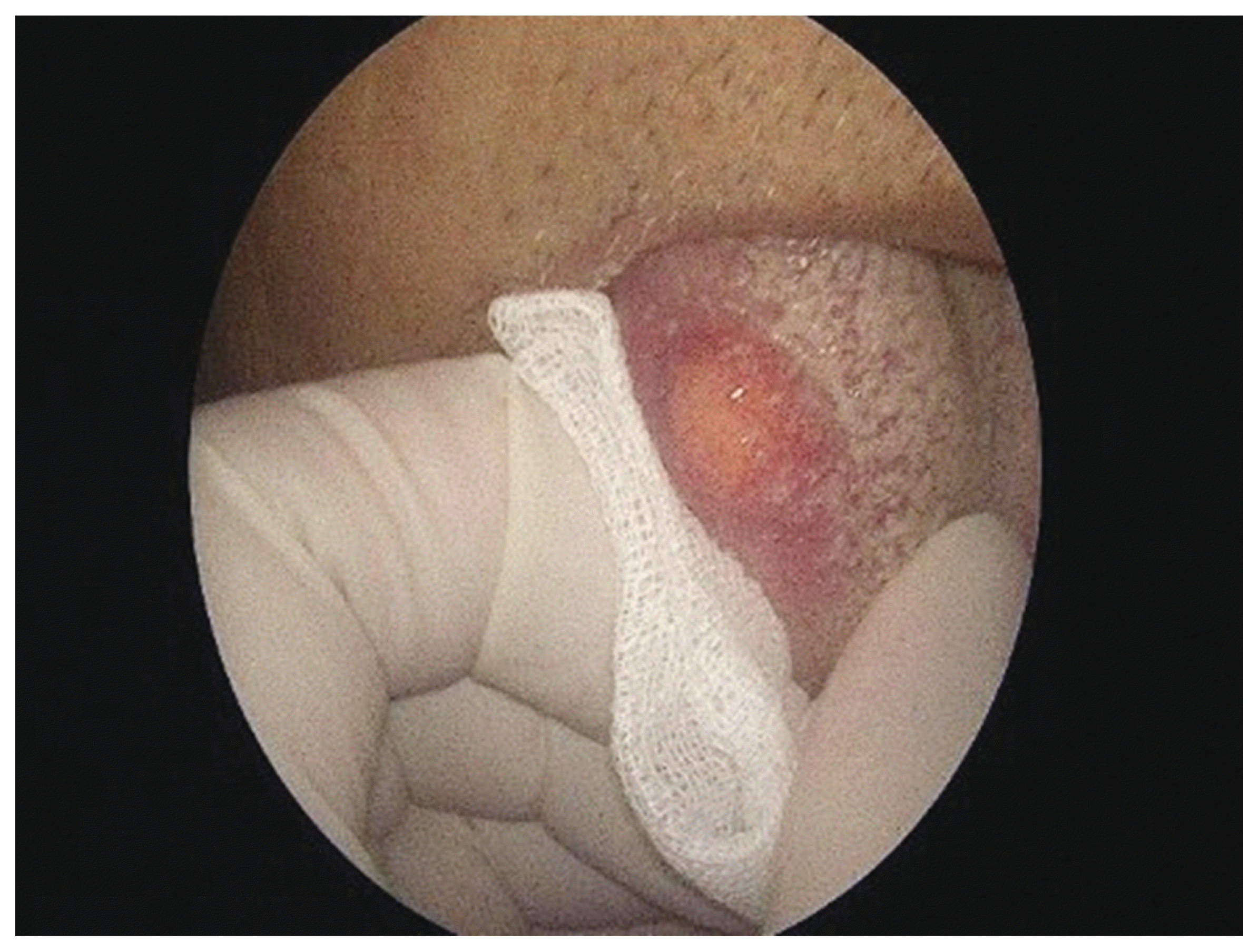
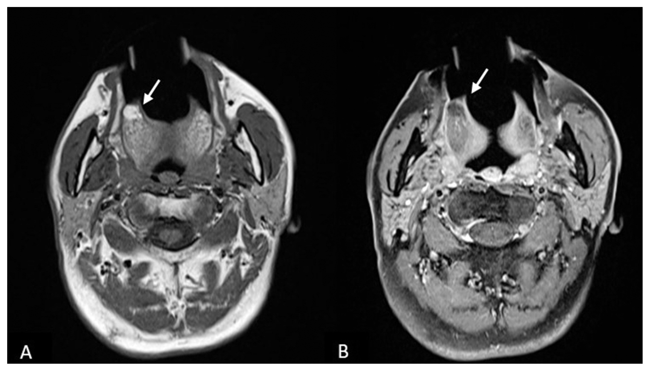
A 76-year-old male patient presented with an asymptomatic, slowly growing tongue mass of unclear onset. The patient had undergone treatment for hypertension, diabetes, and chronic renal failure and was a 30 pack-year ex-smoker. Physical examination revealed a firm cystic mass with a smooth surface, approximately 1 × 1 cm in size, in the right anterior aspect of the tongue (Fig. 1). There was no evidence of cervical lymphadenopathy. Preoperative T1-weighted magnetic resonance imaging (MRI) of the tongue showed a well-defined, ovoid-shaped mass with high-signal intensity, with T1-enhanced MRI showing attenuated signal intensity inside the lesion (Fig. 2). Cervical computed tomography (CT) revealed no cervical lymph node enlargement or other specific findings.
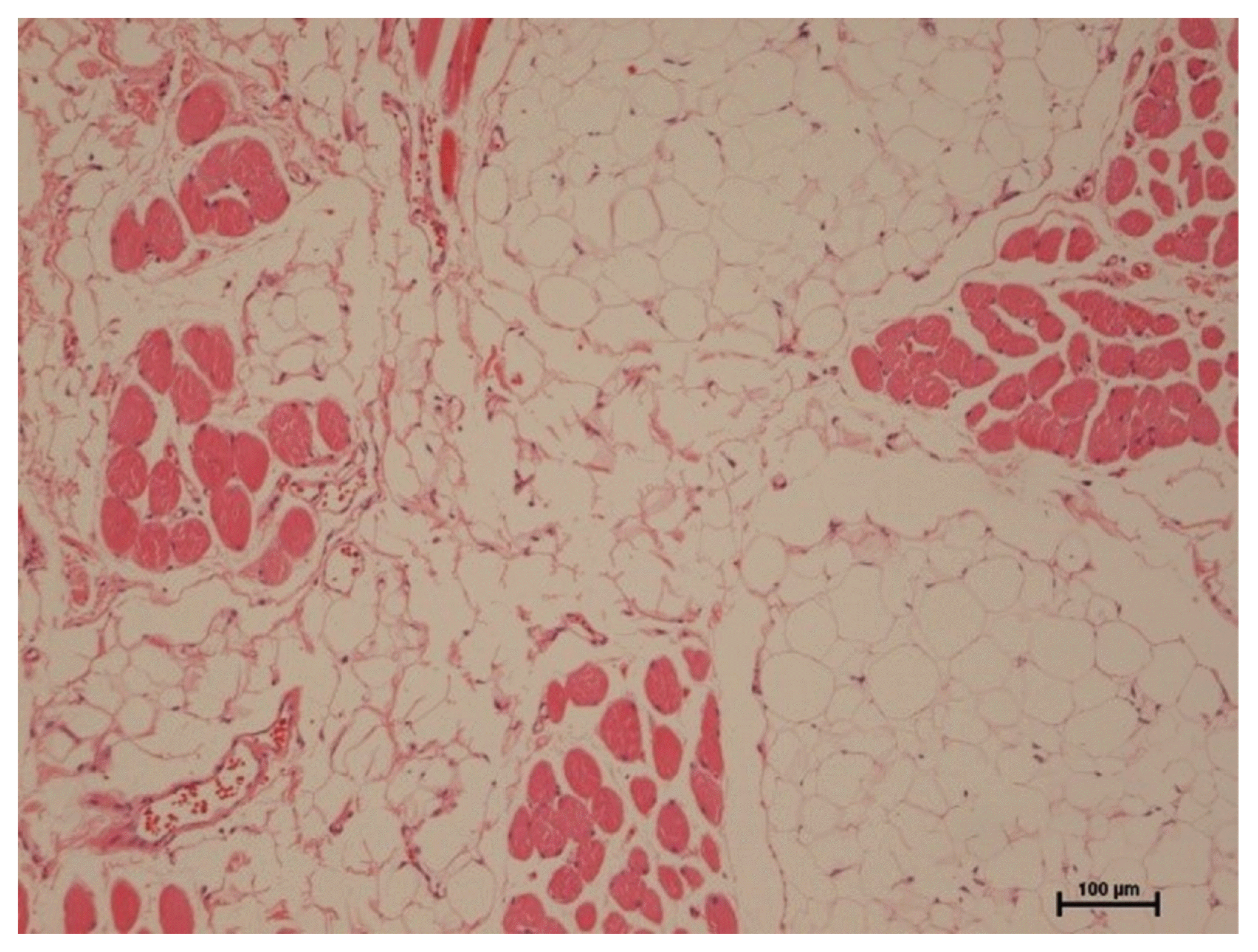
The patient was provisionally diagnosed with a lipoma of the tongue, which was removed surgically. Histopathological examination of the lesion revealed a well-differentiated liposarcoma, also called an atypical lipomatous tumor, with undetermined resection margins. Histologically, the tumor was mainly composed of fatty cells with variation in size and shape and intervening fibrous septum. It also shows characteristic lipoblasts with enlarged indented nuclei and variously sized lipid-containing cytoplasmic vacuoles (Fig. 3).2
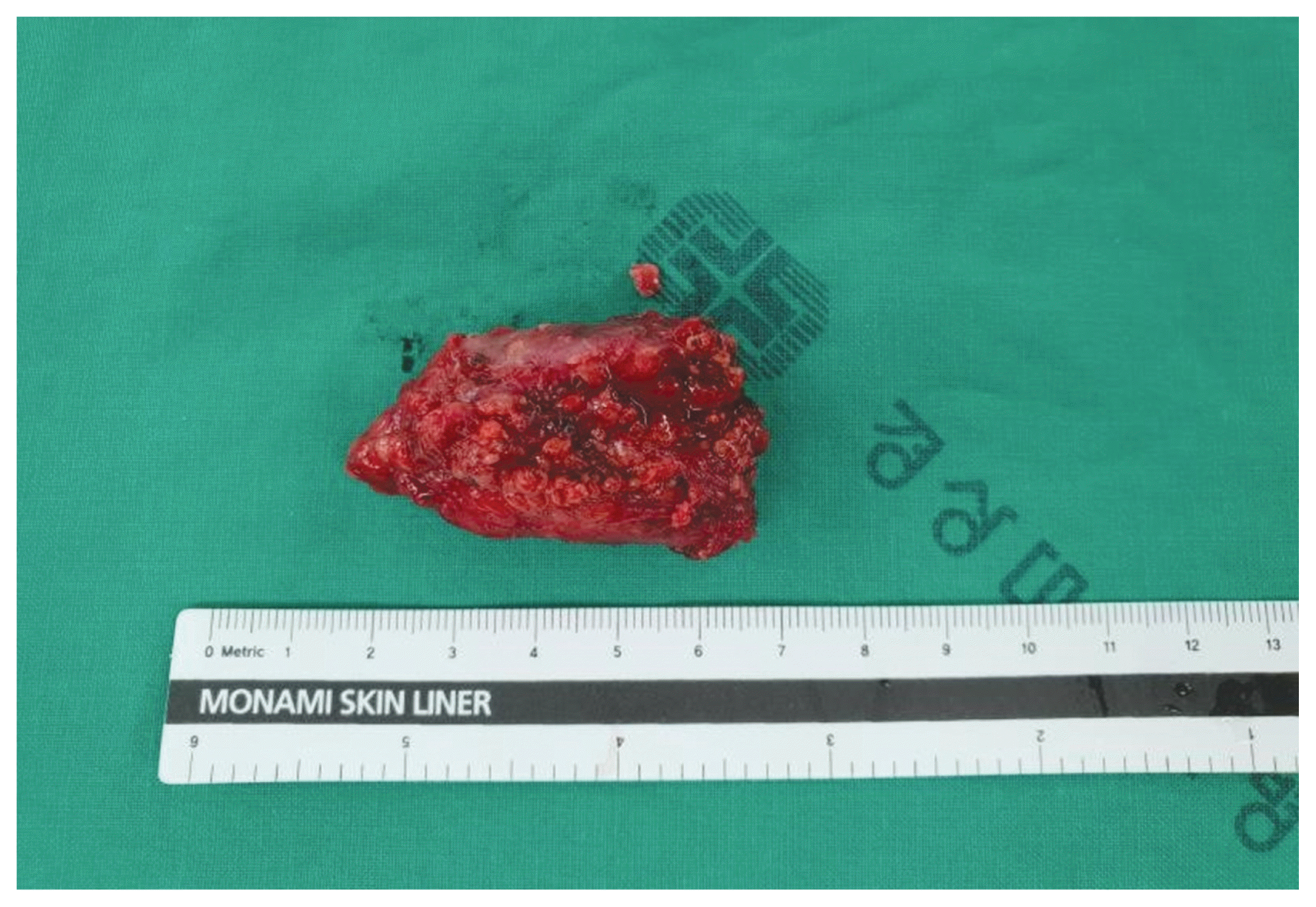
(Fig. 3). The patient was assessed for metastases by positron emission tomography (PET)-CT, abdominal sonography, and gastroscopy, but no evidence of metastasis was observed. A second operation was performed to completely resect the tumor, including safety margins. The postoperative course of the patient was uneventful, with no evidence of tumor recurrence 1 year later.
DISCUSSION
Liposarcomas, first described in 1857, account for approximately 20% of all soft tissue malignancies and are the most common type of sarcoma in adults. Liposarcomas may arise wherever adipose tissue is normally present, but most occur in deep soft tissues and the retroperitoneum. 3,4 Head and neck liposarcomas are uncommon, constituting 1.8 – 6% of all liposarcomas. Oral liposarcomas are extremely rare. The most common sites in the oral cavity for their occurrence are the oral mucosa (38%), the tongue (33%). the floor of the mouth (7%) and the palate (7%).4
Liposarcomas have been classified into several types, including well-differentiated, dedifferentiated, myxoid, pleomorphic and round cell types. Myxoid type is the most common (30 – 50%), followed by well differentiated (20 – 30%), pleomorphic (10 – 25%) and round cell (10 – 15%) types.5 Well-differentiated liposarcomas, especially in the oral cavity, are predominantly superficial, are of small size, and have clear margins. These tumors rarely metastasize or recur. The WHO has classified well-differentiated liposarcomas as atypical lipomatous tumors. 1 The 5-year survival rate of patients with atypical lipomatous tumors was reported to range from 75% to 100%, compared with a 20% 5-year survival rate in patients with pleomorphic and round cell type tumors.6
The distinction between lipoma and atypical lipomatous tumor, however, is a frequent diagnostic dilemma.2 Although these tumors are firmer and less easily compressed and fixed than benign lipomas, they are not easy to distinguish clinically.7 The diagnosis of atypical lipomatous tumors is based on pathologic findings, including the presence of lipoblasts, variations in adipocyte size, and adipocytes with atypical and enlarged nuclei.8,9 Liposarcoma of the tongue can easily be mistaken for benign lipoma, traumatic fibroma, papilloma, generalized gingiva hyperplasia, and pyogenic granuloma.2
Surgical resection is the treatment of choice.7 Wide excision, including safety margins, can significantly reduce recurrence rates.9 However some groups recommended conservative surgical excision with close and long periods of follow–up instead of radical surgical approach due to a generally favorable prognosis based on the histopathologic subtype, location and clear surgical margins.10 Local recurrence rates are reported to range from 7% to 30% due to incomplete resection.4,11 Postoperative radiation therapy may prevent tumor recurrence, especially of radiation sensitive myxoid type tumors. Postoperative radiation therapy is not required for atypical lipomatous tumors if resection margins are tumor free and there is no capsular invasion.12,13 The patient described in this report did not receive additional radiation therapy because there was no involvement of the capsule after wide excision. The average size of liposarcomas of the tongue is 0.8 cm, with prognosis being better in patients with tumors < 5 cm in size.14 By contrast, patients with liposarcomas arising in the retroperitoneum or trunk have a poor prognosis due to the difficulty of surgical resection.4,15
Despite its unclear onset, the tumor in this patient was thought to be benign because of its size and characteristics, including its cystic appearance and smooth surface on palpation. Furthermore, imaging suggested that it was a lipoma. However, histologic examination of the lesion suggested that it was an atypical lipomatous tumor. The diagnosis is made histologically. 12 These tumors should be actively treated if their duration is long or unclear and/or they are large in size. In summary, this report described a patient with a rare atypical lipomatous tumor/well-differentiated liposarcoma of the tongue. The tumor was successfully surgically removed, with no evidence of recurrence at 1-year follow up.
 XML Download