

 PDF
PDF Citation
 Print
Print


Ischemic mitral regurgitation (IMR) has been reported to have chronic, acute, and transient forms. Chronic IMR is generally known as a consequence of left ventricular remodeling and mitral leaflet and chordae tethering. Acute IMR is an uncommon but serious complication of myocardial infarction that is associated with posterior papillary muscle rupture and dysfunction, requiring surgical intervention.1 Acute reversible IMR, although less frequently reported, occurs in patients with normal left ventricular size, systolic function, and mitral valve structure. 1, 2 Further, only a few studies have investigated the importance and treatment of acute transient IMR. Here, we describe a 71-year-old man who had a history of unstable angina, which led to acute mitral regurgitation (MR). The MR was resolved immediately after percutaneous coronary intervention (PCI).
CASE
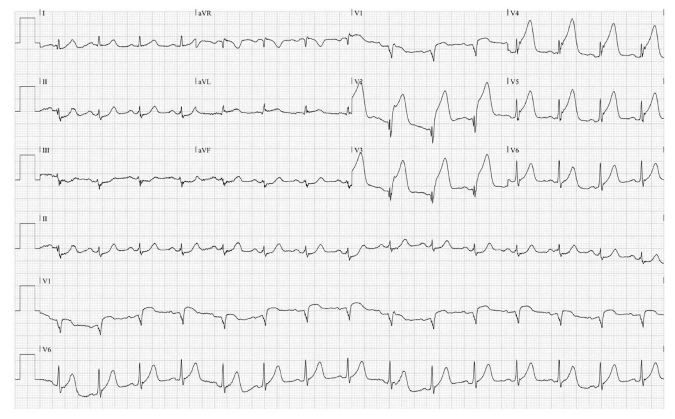
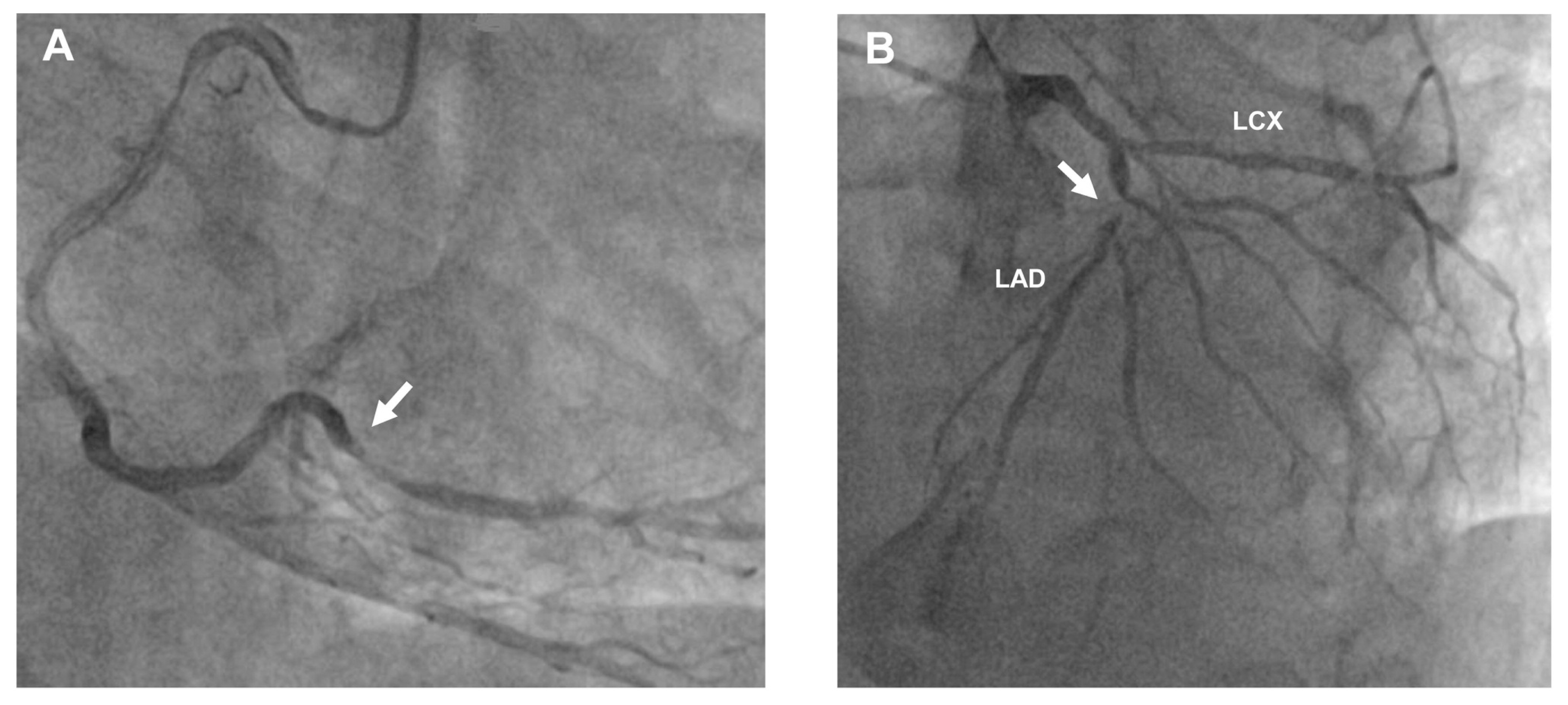
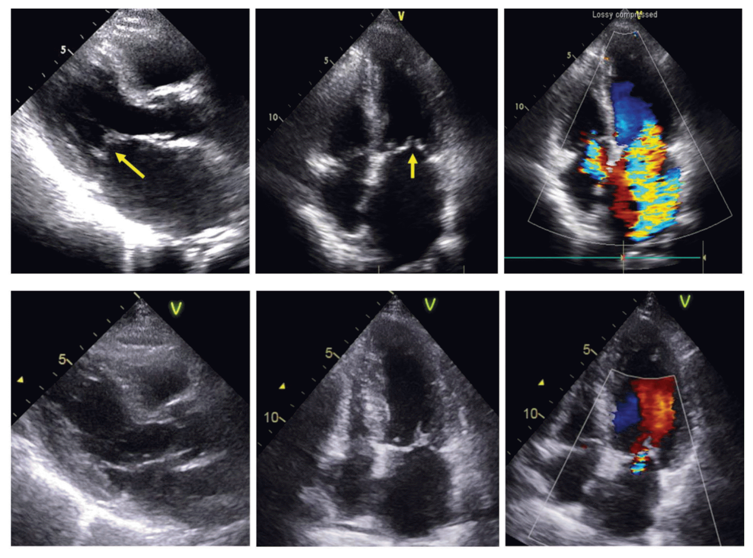
A 71-year-old man with chest pain, which started after exercise, was admitted to the emergency room. He had a history of diabetes mellitus and stable angina with mild left anterior descending artery (LAD) stenosis, which was diagnosed 10 years earlier. Physical examination revealed clear lung sounds without crackles. An initial electrocardiogram showed ST segment elevation in the anterior leads (Fig. 1). Initial blood pressure was 110/70 mmHg, heart rate 74 beats/min, respiratory rate 18 breaths/min, O2 saturation 100% with room air. Primary PCI was performed, and it revealed near-total mid LAD occlusion and about 70% stenosis of the distal right coronary artery (RCA) (Fig. 2). For the culprit coronary artery, mid LAD stenosis, thrombectomy and stent insertion (Synergy 2.5 × 24 mm, Boston Scientific Corporation, Marlborough, MA) were successfully performed. On transthoracic echocardiography (TTE) post-primary PCI, mild MR and hypokinetic motion of the mid to apical portion of the anterior wall were defined. Troponin I was elevated to > 23 ng/mL, creatine kinase-MB (CK-MB) was 300 ng/mL, and N-terminal pro B-type natriuretic peptide (NT-proBNP) level was 78.9 pg/mL. We decided not to perform any more intervention for the RCA and to start the medical therapy, including aspirin, ticagrelor, statin, a beta-blocker, and an angiotensin-receptor blocker. Ten days post-PCI, he presented with chest pain again, but he experienced relief after receiving a tablet of nitroglycerin sublingually. A repeat electrocardiogram showed normal sinus rhythm and no abnormal ST-T change, except the Q wave at the anterior precordial leads. There was no significant change in wall motion abnormality, left ventricular function, and valvular function on the follow-up echocardiography. Initial chest X-ray was clear without pulmonary edema. Treadmill exercise electrocardiography was performed according to the modified Bruce protocol. The test revealed ST segment elevation in aVR at stage 4 and ST segment depression in II, III, and aVF from stage 3 to the recovery phase. He complained of dyspnea immediately after the exercise. Troponin I and CK-MB levels were 0.13 ng/mL and 3.1 ng/mL, respectively. NT-proBNP level was elevated to 1,594 pg/mL. Chest radiography revealed diffuse bilateral infiltration, suggesting pulmonary edema. TTE demonstrated incomplete closure of the mitral valve with severe MR (Fig. 3). No additional wall motion abnormalities were noted; rather, mid-wall motion seemed to be improved. LV size and ejection fraction were within normal ranges. On performing coronary angiography, we confirmed that the previous LAD stent was patent and proceeded with RCA revascularization with stenting. TTE was repeated immediately after PCI and it revealed that the severe MR and incomplete coaptation were resolved (Fig. 3). The patient improved rapidly from a state of acute heart failure.
DISCUSSION
IMR is a frequent complication, and it also has important clinical impacts on the outcomes of ischemic heart disease.2,3 However, its acute transient form has been reported infrequently. Moreover, using a left ventricular angiogram, Finelli et al.4 demonstrated severe transient IMR in a patient with symptoms of recurrent heart failure. After introducing coronary revascularization technique, a few cases of resolution of acute IMR after coronary angioplasty have been reported.5,6 Although the pathogenesis of transient IMR is unclear, posteromedial papillary muscle dysfunction due to ischemia is suggested to be the main causative mechanism. Acute severe IMR in acute myocardial infarction is known as an irreversible mechanical complication caused by papillary muscle rupture and/or dysfunction, which is related to pap50 illary muscle infarction. However, previous reports indicated that there is a transient form of IMR and that this IMR type could disappear after coronary angioplasty.5,6 In our case, the patient with unstable angina who had minimal MR, acute severe MR and pulmonary edema were suddenly developed after treadmill exercise test. Remarkably, the acute severe IMR disappeared immediately after PCI for the RCA. To our knowledge, there is no echocardiographic documentation of abrupt IMR change in a patient with acute coronary syndrome. Recently, transient IMR due to coronary artery spasm was reported and the authors named it the “Eclipsed MR”.1,7,8 Liang et al. suggested that the mechanism behind transient IMR due to spasms is global subendocardial ischemia, which results in apical displacement of both papillary muscles.1 They also explained that the ejection fraction appeared to be preserved because of an acute decrease in afterload caused by severe acute MR. The feature of TTE in our patient was similar to that in global subendocardial ischemia, although the main cause, which is localized posteromedial papillary muscle dysfunction related to RCA stenosis, is different. Therefore, coronary artery disease should be considered in the differential diagnosis of patients with transient IMR even in situations where the mitral valve is structurally normal, wall motion abnormality is uncertain, and ejection fraction is preserved. The treatment of IMR remains inconclusive. In a population of patients with acute myocardial infarction, successful primary PCI was helpful in the improvement of IMR.2,3 In the current case, significant RCA stenosis was recorded in the coronary angiogram for acute anterior ST-elevated myocardial infarction. During the second episode of unstable angina, severe IMR was detected. Because we considered the possibility of papillary muscle dysfunction due to RCA stenosis, early revascularization could be decided. In conclusion, acute transient IMR is an infrequently reported and under-recognized complication. We described a case of acute transient IMR caused by acute coronary syndrome, in which the patient fully recovered immediately after PCI. We suggest that coronary artery disease should be considered in the differential diagnosis of patients with transient IMR. Early diagnosis and prompt therapeutic intervention may help improve the prognosis of these patients.
 XML Download