

 PDF
PDF ePub
ePub Citation
 Print
Print


Dear Editor:
Dermatophytosis is a common skin disease affecting the surface of the skin, including the stratum corneum, hair and nails. However, it is also possible for dermatophytes to invade the dermis and subcutaneous fat layer, resulting in deep dermatophytosis1. Deep infection by dermatophytes is defined as the growth of dermatophytes in the dermis and subcutis, and is believed to result from lesions in superficial dermatophytosis or rupture of infected follicles into the dermis2. The deep lesions are usually accompanied by superficial dermatophytosis3.
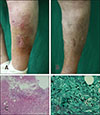
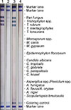
A 78-year-old female presented with reddish patches with purulent discharge on the left lower leg without any trauma for 1 week. She had a history of hypertension, angina and previously undiagnosed onychomycosis, but no other illnesses that would lower immunity. Physical examination showed localized coin-sized erythematous swollen patches with a central ulcer on the left lower leg (Fig. 1A, B). KOH and fungal culture preparation of epidermal scales and purulent discharge were all found to be negative five and two times each, respectively. The histopathologic evaluation demonstrated ill-demarcated, non-encapsulated granulomatous inflammation and an abscess composed of multinucleated giant cells, lymphohistiocytes, and neutrophils in the deep dermis and subcutaneous fat layer. Immunohistochemistry revealed negativity for Gram stain and many acute angle branching hyphae in the deep dermis with periodic acid-Schiff and Gomori Methenamin-silver stain (GMS) (Fig. 1C, D). Trichophyton rubrum was identified by REBA Fungus-ID® (Optipharm M&D, Inc., Wonju, Korea) in biopsy tissue (Fig. 2). Based on clinical and histologic findings, the patient was diagnosed with deep dermatophytosis. We received the patient's consent form about publishing all photographic materials.
The patient was initially treated with oral ciprofloxacin for 4 weeks and terbinafine for 10 weeks. After 14 weeks with clinical improvement, many hyphae were still observed on follow-up biopsy of the deep dermis with GMS stain, and we replaced terbinafine with oral fluconazole for 12 weeks. After 3 months, the skin lesion was completely resolved, and there was no evidence of new lesions. Deep dermatophytosis is a rare disease, with few prior reported cases. In a recent article, Kim et al.3 provided a descriptive review of 46 cases of deep dermatophytosis, of which 38 (83%) were immunocompromised. Most patients had pre-existing superficial dermatophytosis. Kershenovich et al.4 reported 10 cases of deep dermatophytosis. In all cases, a concurrent superficial dermatophyte infection was documented, and all patients were immunocompromised. Because deep dermatophytosis in immunocompetent patients is very rarely reported35, we did not suspect the condition at first.
Kershenovich et al.4 reported that terbinafine was effective in the treatment of deep dermatophytosis, and the average duration of treatment with terbinafine was 7 weeks (range, 4~12 weeks). However, despite sufficient use of terbinafine, the treatment was not effective in our patient. Therefore, we decided to replace it with fluconazole, a good treatment option for deep dermatophytosis.
We report a rare case of dermal infection of T. rubrum in an immunocompetent patient. Even if patients are immunocompetent, clinicians need to consider the possibility of deep dermatophytosis with untreated ulcerative lesions. A history of superficial dermatophytosis and skin biopsy can be helpful to confirm the diagnosis.
 XML Download