



 PDF
PDF ePub
ePub Citation
 Print
Print


Hyoungyoung Kim, Jeongmin Choi
Figures and Tables
Fig. 1
(A) Upper endoscopy showed 1 cm sized subepithelial lesion with central depression was located at the antrum. (B) Endoscopic ultrasonography showed ill-defined hypoechoic lesion located in the submucosal layer (arrow).
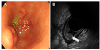
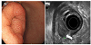
Fig. 2
(A) Upper endoscopy showed semi-pedunculated, 3 cm sized subepithelial mass with central depression was located at the antrum. (B) Endoscopic ultrasonography showed hypoechoic mass originating from the muscularis mucosa (arrow).
References
1. Liu TC, Lin MT, Montgomery EA, Singhi AD. Inflammatory fibroid polyps of the gastrointestinal tract: spectrum of clinical, morphologic, and immunohistochemistry features. Am J Surg Pathol. 2013; 37:586–592.
2. Albuquerque A, Rios E, Carneiro F, Macedo G. Evaluation of clinico-pathological features and Helicobacter pylori infection in gastric inflammatory fibroid polyps. Virchows Arch. 2014; 465:643–647.

3. Kang HC, Menias CO, Gaballah AH, et al. Beyond the GIST: mesenchymal tumors of the stomach. Radiographics. 2013; 33:1673–1690.
4. Miyata T, Yamamoto H, Kita H, et al. A case of inflammatory fibroid polyp causing small-bowel intussusception in which retrograde double-balloon enteroscopy was useful for the preoperative diagnosis. Endoscopy. 2004; 36:344–347.
5. Hizawa K, Iida M, Tada S, et al. Endoscopic evaluation of gastric inflammatory fibroid polyp. Surg Endosc. 1995; 9:397–400.
6. Matsushita M, Uchida K, Nishio A, Okazaki K. Endoscopic and EUS features of gastric inflammatory fibroid polyps. Gastrointest Endosc. 2009; 69:188. author reply 188-189.
7. Pantanowitz L, Antonioli DA, Pinkus GS, Shahsafaei A, Odze RD. Inflammatory fibroid polyps of the gastrointestinal tract: evidence for a dendritic cell origin. Am J Surg Pathol. 2004; 28:107–114.
8. Mucientes P, Mucientes F, Klaassen R. Inflammatory fibroid polyp associated with early gastric carcinoma: a case report. Ann Diagn Pathol. 2012; 16:148–151.
9. Kim HS, Kim JI, Koh MB, et al. Gastric inflammatory fibroid polyp resected by endoscopic submucosal dissection. Korean J Gastrointest Endosc. 2009; 38:224–228.
10. Kim YI, Kim WH. Inflammatory fibroid polyps of gastrointestinal tract: evolution of histologic patterns. Am J Clin Pathol. 1988; 89:721–727.
11. Kim HC, Yang DM, Jin W, Nam DH, Kim GY, Choi SI. CT findings of a gastric inflammatory fibroid polyp manifesting as unexpected growth: a case report. J Korean Soc Radiol. 2009; 60:271–274.
12. Kim KH, Seo HJ, Kim JI, et al. Inflammatory fibroid polyp of the stomach mimicking gastric cancer. J Korean Surg Soc. 2003; 65:72–75.
13. Lee JH, Yoo JS, Jung HY, et al. A case of invasion of muscularis propria of gastric inflammatory fibroid polyp. Korean J Helicobacter Up Gastrointest Res. 2015; 15:254–257.
14. Cho NH, Jeong HJ, Kim HG. A pathological and immunohistochemical study of 9 cases of inflammatory fibroid polyp. Korean J Pathol. 1989; 23:20–28.
15. Han GJ, Kim JH, Lee SS, Park SH, Lee JS, Ha HK. Inflammatory fibroid polyps of the gastrointestinal tract: a 14-year CT study at a single institution. Abdom Imaging. 2015; 40:2159–2166.
16. Kim MK, Higgins J, Cho EY, Ko YH, Oh YL. Expression of CD34, bcl-2, and kit in inflammatory fibroid polyps of the gastrointestinal tract. Appl Immunohistochem Mol Morphol. 2000; 8:147–153.
17. Harima H, Kimura T, Hamabe K, et al. Invasive inflammatory fibroid polyp of the stomach: a case report and literature review. BMC Gastroenterol. 2018; 18:74.
18. Zinkiewicz K, Zgodzinski W, Dabrowski A, Szumilo J, Cwik G, Wallner G. Recurrent inflammatory fibroid polyp of cardia: a case report. World J Gastroenterol. 2004; 10:767–768.
 XML Download