



 PDF
 Citation
 Print


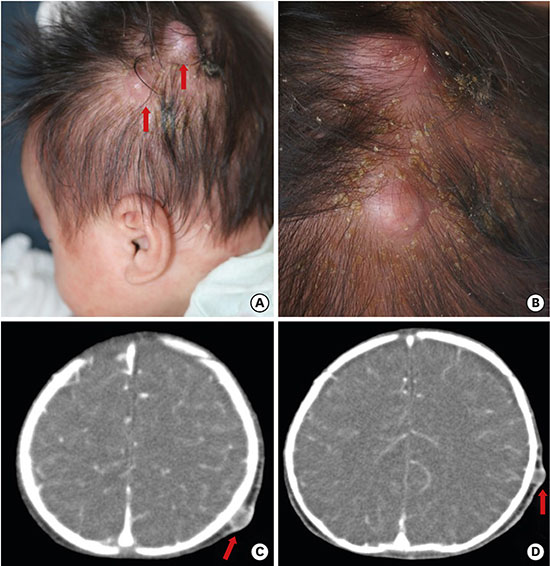
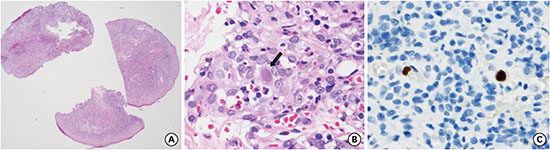
A seven-week-old male baby was admitted to the hospital with scaly patches and nodules with discharge. He was born at term with an uncomplicated perinatal course and a weight of 3.1 kg. The baby was formula-fed after birth and showed normal growth. At approximately three weeks after delivery, he developed a vesicular rash; the lesion progressed to oozing and crusting and was diagnosed as “seborrheic dermatitis.” The symptoms initially improved with the use of a topical steroid; however, they deteriorated in the week before admission. Skin examination revealed two erythematous bean-sized nodules with yellowish oozing and crusting and diffuses scaly patches on his scalp (Fig. 1A and B). The rest of the skin examination was normal. A general physical examination revealed splenomegaly but no other specific finding. A computed tomographic scan showed several peripheral-enhancing, well-defined cystic lesions in the subcutaneous layer of his scalp, which suggested an epidermal cyst with inflammatory change (Fig. 1C and D). The dermatologist assumed seborrheic dermatitis or furunculosis. A 3-mm skin-punch biopsy was performed from a nodule on his scalp and the bacterial culture was done. Microscopically, the section revealed an ulcerative lesion composed of mainly granulation tissue admixed with acute and chronic inflammatory cells (Fig. 2A). Several typical eosinophilic intranuclear inclusions were noted in the endothelial cells of the blood vessels (Fig. 2B). They were immunopositive for cytomegalovirus (CMV) antibody, which was consistent with CMV infection (Fig. 2C). After a skin biopsy, we finally diagnosed the infant with cutaneous CMV infection. CMV immunoglobulin M antibody was positive and CMV DNA was detected by polymerase chain reaction in his blood and urine. Brain sonography showed no intracranial abnormality; and ophthalmologic examination showed no evidence of retinal involvement. An otoacoustic emissions test revealed no hearing deficit. Assessments for possible immunodeficiency including human immunodeficiency virus serology, immunoglobulin class, T- and B-cell subsets, complement components, and neutrophil oxidative burst assay were normal. There was no abscess formation at bacillus Calmette-Guérin (BCG) site and no familial history of immunodeficiency. The lesions gradually resolved without CMV-specific therapy. Follow-up at 2 years of age, patients showed normal growth and no history of significant infections.
Infection with CMV is ubiquitous, and up to 38% of neonates may become infected with various symptoms and signs.1 Cutaneous CMV infection is a rare and, when reported, is mostly revealed in patients who have acquired immune deficiency syndrome or are immunocompromised.23 Skin involvement of CMV may show nonspecific and specific lesions. The nonspecific lesions are mostly due to immunological alterations that follow a viral infection or to hypersensitivity manifestations. Nonspecific lesions are various rashes and eruptions such as macular and papular rashes and urticarial and scarlatiniform eruptions.4 The specific lesions differ according to host immunity. In immunocompromised hosts, CMV infection shows a wide spectrum of cutaneous manifestation and can signify disseminated infection.5 However, immunocompetent hosts very rarely present with cutaneous CMV infection include purpuric papules and plaque,6 ulcer.7 In the present case, the patient had pruritic, scaly patches and nodules on his face and scalp. Thus, the present case highlights the importance of considering CMV infection in the differential diagnosis of scalp nodule in healthy infants.

 XML Download