

 PDF
PDF ePub
ePub Citation
 Print
Print


Carpal tunnel syndrome is the most common compressive neuropathy of the upper extremity1. Moreover, several causative factors increase the carpal tunnel pressure, which include space-occupying mass, tenosynovitis, aberrant musculature, or congenital abnormal median nerve. A persistent median artery is a numerous reported anatomical variant, which is supposed to degenerate over time23. In addition, studies have shown its co-existence with bifid median nerve. Most cases of persistent median artery are known to be asymptomatic4, although some reports demonstrated that co-existence of persistent median artery with bifid median nerve can lead to carpal tunnel syndrome5. However, a case of thrombosed persistent median artery with bifid median nerve is extremely rare. Hence, we report an unusual case and surgical outcome of thrombosed persistent median artery with bifid median nerve causing acute carpal tunnel syndrome. Informed consent was obtained from the patient for purpose of the case report.
CASE REPORT
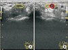
A 58-year-old female patient was referred to corresponding author's hospital for pain at the volar aspect of the left wrist and paresthesia with sensory loss at the median nerve territory from the thumb to the middle finger. The patient had no prior history of taking anti-diabetic, anti-hypertensive, and any thrombogenic-related medications. The chief complaint was pain that occurred acutely, and the symptoms persisted for five weeks. The patient's numbness was not relieved by oral medication. On physical examination, both Tinnel sign and Phalen test were positive. However, no thenar muscle atrophy was noted. Although the electrodiagnostic study at corresponding author's hospital revealed no specific abnormality, we were able to identify a persistent median artery between the divided median nerves on Doppler ultrasound examination (Fig. 1). The persistent median artery was completely obstructed in the carpal tunnel area, but blood flow was identified to be proximal to the carpal tunnel. In addition, we identified a thrombus — that was approximately 4 cm in length — of the persistent median artery at the wrist on both the magnetic resonance image and Doppler ultrasound (Fig. 2). The surgery was performed with an operating microscope. First, we performed open carpal tunnel release. The skin was incised using a curved longitudinal incision, passing the wrist crease. Subsequently, we could identify the split median nerve from the inlet of the carpal tunnel, which is proximally 2 cm. In addition, the radial-side branch of the median nerve was more dominant than the other side, and the persistent median artery was found between the split median nerve. The persistent median artery was completely obliterated in 5 mm diameter, and a 2-cm length of the artery was obliterated proximally and distally from the carpal tunnel inlet. Under the operating microscope, the obstructed artery was resected and ligated proximally (Fig. 3). The patient had previous paresthesia of the affected hand, which was resolved immediately postoperatively, and preoperative chief complaint was completely resolved at 9 months postoperatively.
DISCUSSION
Carpal tunnel syndrome has typical symptoms, including paresthesia or numbness in the region of median nerve distribution1. However, patients typically do not complain about symptoms on the volar aspect of the wrist. If the patient has these symptoms, we should rule out other uncommon causes of compressive neuropathy of the wrist. Acute onset of median nerve entrapment symptom tends to result from trauma, swelling, infections, inflammation, anomalous anatomy, coagulopathy, or tumorous conditions6. In this present case, atypical symptoms were noted, which were painful sensation at the volar aspect of the wrist and abrupt symptom onset. These are the reasons why we performed further evaluation for the possibility of abnormal anatomy or mass-like lesion.
The incidence of bifid median nerve varies from 0.8% to 21.0% in patients with carpal tunnel syndrome7. The various differences of the incidence maybe because of various diagnostic tools or ethnic groups. In addition, several reports have suggested that the bifid median nerve can be a causative factor for carpal tunnel syndrome8. One of the hypotheses is that the bifid median nerve may facilitate compression of the nerve in the carpal tunnel because of larger summated cross-sectional area compared with the cross-sectional area of a non-bifid median nerve8. In contrast, some authors recently reported that the bifid median nerve is not an independent risk factor for development of carpal tunnel syndrome79.
Meanwhile, co-existence of the bifid median nerve and persistent median artery was suggested by several authors29. Embryologically, the upper limb develops at the end of four weeks to eight weeks gestation. Angiogenesis of the upper limb is followed with development of the upper limb. Briefly, the median artery is the main branch of the brachial artery at the forearm area at first, followed by the ulnar and radial arteries that play a major role for providing the blood supply below the forearm level while the median artery degenerates2. At this time, the persistent median artery can cause splitting of the median nerve, which is thus called bifid median nerve. Therefore, when we encounter either the bifid median nerve or persistent median artery, we should evaluate for the cooccurrence of both anatomical abnormalies. Generally, two imaging modalities can identify an abnormal finding in the carpal tunnel, the Doppler ultrasound and magnetic resonance imaging. Most of the cited reports have used a Doppler ultrasound for diagnosis of persistent median artery and bifid median nerve in the carpal tunnel. Based on our present case, we also recommend that the Doppler ultrasound examination is useful for identification of this abnormal anatomy in the carpal tunnel area in the outpatient clinic. In addition, a clear understanding of the abnormal anatomy is very important when performing surgery for carpal tunnel syndrome.
The management of thrombosed persistent median artery with bifid median nerve causing acute carpal tunnel syndrome can be conservative or surgical treatment. Kele et al.5 reported that surgical management of this case produced a good outcome. In contrast, Salter et al.10 considered treatment with anticoagulants as a conservative management. Although he did not mention the accurate period of symptom duration, the report suggested early detection of the thrombosed median artery can be of great help for preventing the need for any surgery or thrombolytics. Our present case also demonstrated an acute onset of compressive median nerve neuropathy, but the symptoms of obliterated median artery had persisted for five weeks. That is why we considered surgery for the management of the obstructed median artery and compressive median nerve neuropathy. Based on our experiences, we think that a problematic vessel in the past can also tend to be problematic in the future, and the carpal tunnel space and proximal area are sites vulnerable to compression of the contents in the carpal tunnel space anatomically. The persistent median artery can also be easily compressed around the carpal tunnel. Hence, we recommend surgical management for this abnormal finding rather than a conservative treatment in the same case, although immediate medical therapy can help avoid surgery in the early stage.
In conclusion, careful identification of an abnormal anatomy in the carpal tunnel is essential, particularly in cases wherein carpal tunnel syndrome occurred abruptly. Among the diagnostic tools, Doppler ultrasound examination is very useful in an outpatient clinic, because it provides a quicker and more accurate diagnosis than magnetic resonance imaging. In addition, we suggest that surgical treatment for a thrombosed persistent median artery in carpal tunnel syndrome provides a good result.


 XML Download