

 PDF
PDF ePub
ePub Citation
 Print
Print


Abstract
Purpose:
The object of this study is to evaluate the clinical and radiographic outcomes of two different kinds of dorsal plating for dorsally displaced distal radius fractures.
Methods:
Forty-three patients with dorsal plate fixation for distal radius fracture were retrospectively reviewed. Twenty-four patients were treated with nonlocking dorsal plate (group I) and nineteen patients with locking dorsal plate (group II). Range of motion and Green and O'Brien score were evaluated as clinical results, and radial inclination, radial length, volar tilt at postoperative and last follow up were evaluated as radiographic results.
Results:
According to AO classification system, there were 18 type A fractures, 4 type B fractures, 21 type C fractures. In group I, the mean flexion and extension were 65.0° and 65.3°, respectively; ulnar and radial deviation were 25.5° and 20.8°; pronation and supination were 80.0° and 80.4°. In group II, the mean flexion and extension were 64.5° and 67.3°, respectively; ulnar and radial deviation were 30.6° and 20.6°; pronation and supination were 81.4° and 78.6°. The mean Green and O'Brien score was 94.4 in group I and 92.2 in group II, and 41 patients had satisfactory result. There was no statistical significant difference in clinical results between the groups. Results of postoperative and last follow-up radiographic analyses for mean radial inclination were 23.9° and 24.1° in group I, respectively, and 24.2° and 24.9° in group II; radial length were 9.9 mm and 9.7 mm in group I, and 10.1 mm and 9.2 mm in group II; mean volar tilt were 12.2° and 13.1° in group I, and 14.8° and 13.7° in group II. There were no statistical significant changes within radiographic parameters. No extensor tendon rupture was reported.
References
1. Graff S, Jupiter J. Fracture of the distal radius: classification of treatment and indications for external fixation. Injury. 1994; 25(Suppl 4):S-D14–25.

2. Benson EC, DeCarvalho A, Mikola EA, Veitch JM, Moneim MS. Two potential causes of EPL rupture after distal radius volar plate fixation. Clin Orthop Relat Res. 2006; 451:218–22.
3. Kamath AF, Zurakowski D, Day CS. Low-profile dorsal plating for dorsally angulated distal radius fractures: an outcomes study. J Hand Surg Am. 2006; 31:1061–7.
4. Yu YR, Makhni MC, Tabrizi S, Rozental TD, Mundanthanam G, Day CS. Complications of low-profile dorsal versus volar locking plates in the distal radius: a comparative study. J Hand Surg Am. 2011; 36:1135–41.
5. Osada D, Tamai K, Iwamoto A, Fujita S, Saotome K. Dorsal plating for comminuted intra-articular fractures of the distal end of the radius. Hand Surg. 2004; 9:181–90.
6. Axelrod TS, McMurtry RY. Open reduction and internal fixation of comminuted, intraarticular fractures of the distal radius. J Hand Surg Am. 1990; 15:1–11.
7. Ring D, Jupiter JB, Brennwald J, Buchler U, Hastings H 2nd. Prospective multicenter trial of a plate for dorsal fixation of distal radius fractures. J Hand Surg Am. 1997; 22:777–84.
8. Carter PR, Frederick HA, Laseter GF. Open reduction and internal fixation of unstable distal radius fractures with a low-profile plate: a multicenter study of 73 fractures. J Hand Surg Am. 1998; 23:300–7.
9. Jupiter JB. Plate fixation of fractures of the distal aspect of the radius: relative indications. J Orthop Trauma. 1999; 13:559–69.
10. Jupiter JB, Fernandez DL. Complications following distal radial fractures. Instr Course Lect. 2002; 51:203–19.
11. Campbell DA. Open reduction and internal fixation of intra articular and unstable fractures of the distal radius using the AO distal radius plate. J Hand Surg Br. 2000; 25:528–34.
12. Chiang PP, Roach S, Baratz ME. Failure of a retinacular flap to prevent dorsal wrist pain after titanium Pi plate fixation of distal radius fractures. J Hand Surg Am. 2002; 27:724–8.
13. Kambouroglou GK, Axelrod TS. Complications of the AO/ASIF titanium distal radius plate system (pi plate) in internal fixation of the distal radius: a brief report. J Hand Surg Am. 1998; 23:737–41.
14. Orbay JL, Fernandez DL. Volar fixation for dorsally displaced fractures of the distal radius: a preliminary report. J Hand Surg Am. 2002; 27:205–15.
15. Musgrave DS, Idler RS. Volar fixation of dorsally displaced distal radius fractures using the 2.4-mm locking compression plates. J Hand Surg Am. 2005; 30:743–9.
16. Haug LC, Glodny B, Deml C, Lutz M, Attal R. A new radiological method to detect dorsally penetrating screws when using volar locking plates in distal radial fractures. The dorsal horizon view. Bone Joint J. 2013; 95B:1101–5.
17. Lee SK, Bae KW, Choy WS. Use of the radial groove view intra-operatively to prevent damage to the extensor pollicis longus tendon by protruding screws during volar plating of a distal radial fracture. Bone Joint J. 2013; 95B:1372–6.
18. Simic PM, Robison J, Gardner MJ, Gelberman RH, Weiland AJ, Boyer MI. Treatment of distal radius fractures with a low-profile dorsal plating system: an outcomes assessment. J Hand Surg Am. 2006; 31:382–6.
19. Lutsky K, Boyer M, Goldfarb C. Dorsal locked plate fixation of distal radius fractures. J Hand Surg Am. 2013; 38:1414–22.
Fig. 1.
Dorsal approach for distal radius fracture. (A) Skin incision over Lister's tubercle with zigzag pattern at dorsal aspect of wrist joint. (B) Subperiosteal elevation after dissection of extensor retinaculum. (C) Dorsal plate fixation after the reduction of fracture site and temporary fixation with K-wires. (D) Proximal half of extensor retinaculum was used to cover plate and distal screws. (E) Distal half of extensor retinaculum was repaired. (F) Extensor pollicis longus tendon was placed as subcutaneous.
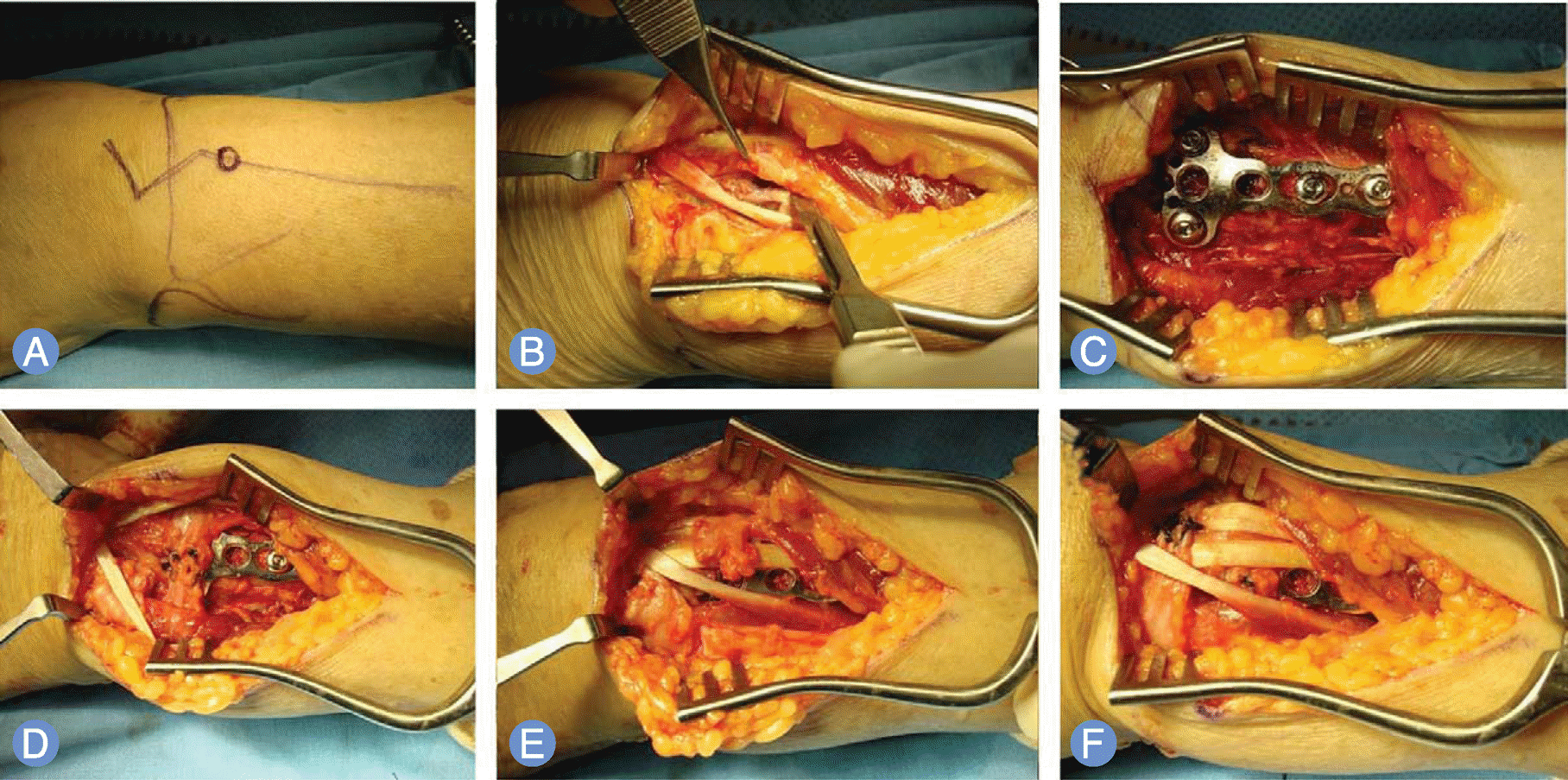
Fig. 2.
Sixty-eight-year-old male, with dorsally displaced unstable distal radius fracture, was treated with open reduction and internal fixation using dorsal locking T-plate. (A) Preoperative radiographs show AO A3 type distal radius fracture. (B) Postoperative radiographs show good reduction and fixation. (C) Twelve months later, radiographs show no interval change.
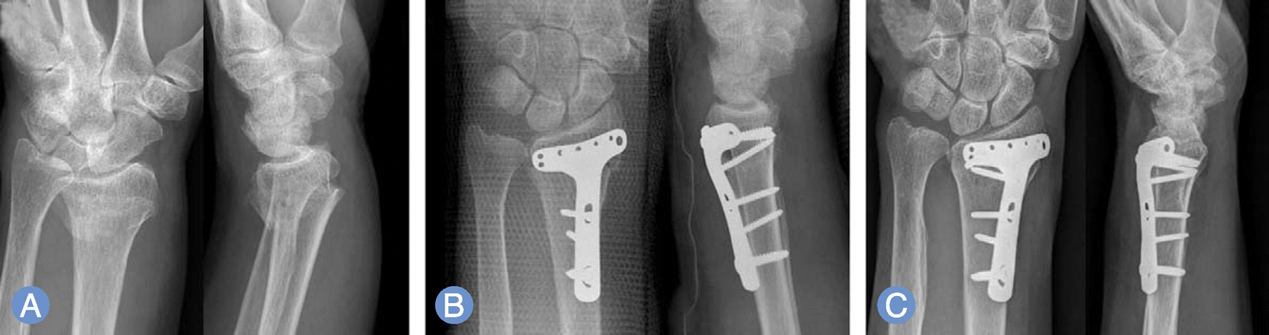
Table 1.
Range of motion
| Type of motion | Total | Group I | Group II | p-value |
|---|---|---|---|---|
| Flexion (°) | 65.8 (25–90) | 65.0 (25–90) | 64.5 (40–90) | 0.958* |
| Extension (°) | 66.2 (30–90) | 65.3 (30–90( | 67.3 (30–90) | 0.825* |
| Ulnar deviation (°) | 27.4 (0–45) | 25.5 (0–35) | 30.6 (15–45) | 0.515* |
| Radial deviation (°) | 20.7 (5–40) | 20.8 (5–40) | 20.6 (5–35) | 1.0* |
| Pronation (°) | 80.7 (40–90) | 80.0 (40–90) | 81.4 (50–90) | 0.837* |
| Supination (°) | 79.3 (40–90) | 80.4 (40–90) | 78.6 (40–90) | 0.650* |
Table 2.
Functional outcomes
| Green and O'Brien score | Total | Group I | Group II | p-value |
|---|---|---|---|---|
| Pain (0-25) | 21.5 (15–25) | 20.8 (15–25) | 22.3 (15–25 | - |
| Range of motion (5-25) | 21.4 (10–25) | 21.4 (10–25) | 21.4 (15–25) | - |
| Occupation (0–25) | 19.7 (5–25) | 21.1 (15–25) | 17.7 (5–25) | - |
| Grip power (0–10) | 6.8 (5–10) | 6.7 (5–10) | 6.9 (5–10 | - |
| X-ray (0–25) | 24.3 (20–25) | 24.4 (20–25) | 24.1 (20–25) | - |
| Total | 94.0 (70–110) | 94.4 (70-110) | 92.2 (75–110) | 0.659* |
Table 3.
Changes in radiographic parameters
| Radial inclination (°) | Radial length (mm) | Volar tilt (°) | |
|---|---|---|---|
| Total | |||
| Postoperative | 24.0±4.22 | 10.0±2.44 | 13.2±8.2 |
| Latest follow-up | 24.4±4.04 | 9.5±3.0 | 13.3±7.7 |
| p-value | 0.523* | 0.091* | 0.930* |
| Group I | |||
| Postoperative | 23.9±4.50 | ± | 12.2±8.47 |
| Latest follow-up | 24.1 ±4.70 | ± | 13.1±7.63 |
| p-value | 0.801* | 0.528* | 0.248* |
| Group II | |||
| Postoperative | 24.2±3.97 | 10.1±2.45 | 14.8±7.89 |
| Latest follow-up | 24.9±2.90 | 9.2±1.70 | 13.7±8.15 |
| p-value | 0.479* | 0.084* | 0.844† |
 XML Download