

 PDF
PDF ePub
ePub Citation
 Print
Print


Abstract
Purpose:
The purpose of this study is to compare the clinical results between two different methods of hindfoot endoscopy to treat posterior ankle impingement syndrome.
Materials and Methods:
Between January 2008 and January 2014, 52 patients who underwent hindfoot endoscopy were retrospectively reviewed. Two methods of hindfoot endoscopy were used; Group A was treated according to van Dijk and colleagues’ standard two-portal method, and group B was treated via the modified version of the above, using a protection cannula. For clinical comparison, the American Orthopaedic Foot and Ankle Society (AOFAS) hindfoot score, time required to return to activity, and the presence of complications were used.
Results:
There was no statistically significant difference in the AOFAS scores at the final follow-up, and there was also no statistically significant difference in the times for the scores to return to the preoperative level. There were no permanent neurovascular injuries and wound problems in either group.
Conclusion:
Use of protection cannula may provide additional safety during hindfoot endoscopy. We could not prove whether protection cannula can provide superior safety for possible neurovascular injury. Considering the possible safety and risk of using additional instrument, the use of this method may be optional.
REFERENCES
1.Stone JW., Guhl JF. Diagnostic arthroscopy of the ankle. Andrews JR, Tinnerman LA, editors. editors.Diagnostic and operative arthroscopy. Philadelphia: WB Saunders;1997. p. 423–30.
2.Jerosch J., Fadel M. Endoscopic resection of a symptomatic os trigonum. Knee Surg Sports Traumatol Arthrosc. 2006. 14:1188–93.

3.Golanó P., Vega J., Pérez-Carro L., Götzens V. Ankle anatomy for the arthroscopist. Part I: the portals. Foot Ankle Clin. 2006. 11:253–73.
4.Ferkel RD., Scranton PE Jr. Arthroscopy of the ankle and foot. J Bone Joint Surg Am. 1993. 75:1233–42.
5.Ferkel RD., Heath DD., Guhl JF. Neurological complications of ankle arthroscopy. Arthroscopy. 1996. 12:200–8.
6.van Dijk CN., Scholten PE., Krips R. A 2-portal endoscopic approach for diagnosis and treatment of posterior ankle pathology. Arthroscopy. 2000. 16:871–6.
7.Abramowitz Y., Wollstein R., Barzilay Y., London E., Matan Y., Sha-bat S, et al. Outcome of resection of a symptomatic os trigonum. J Bone Joint Surg Am. 2003. 85:1051–7.
8.Guo QW., Hu YL., Jiao C., Ao YF., Tian DX. Open versus endoscopic excision of a symptomatic os trigonum: a comparative study of 41 cases. Arthroscopy. 2010. 26:384–90.
9.van Dijk CN. Hindfoot endoscopy. Foot Ankle Clin. 2006. 11:391–414.
10.van Dijk CN. Hindfoot endoscopy for posterior ankle pain. Instr Course Lect. 2006. 55:545–54.
11.Acevedo JI., Busch MT., Ganey TM., Hutton WC., Ogden JA. Coaxial portals for posterior ankle arthroscopy: an anatomic study with clinical correlation on 29 patients. Arthroscopy. 2000. 16:836–42.
Figure 1.
Simple lateral radiograph of ankle revealed osteophyte of posterior aspect of the talus on lateral view (arrow).
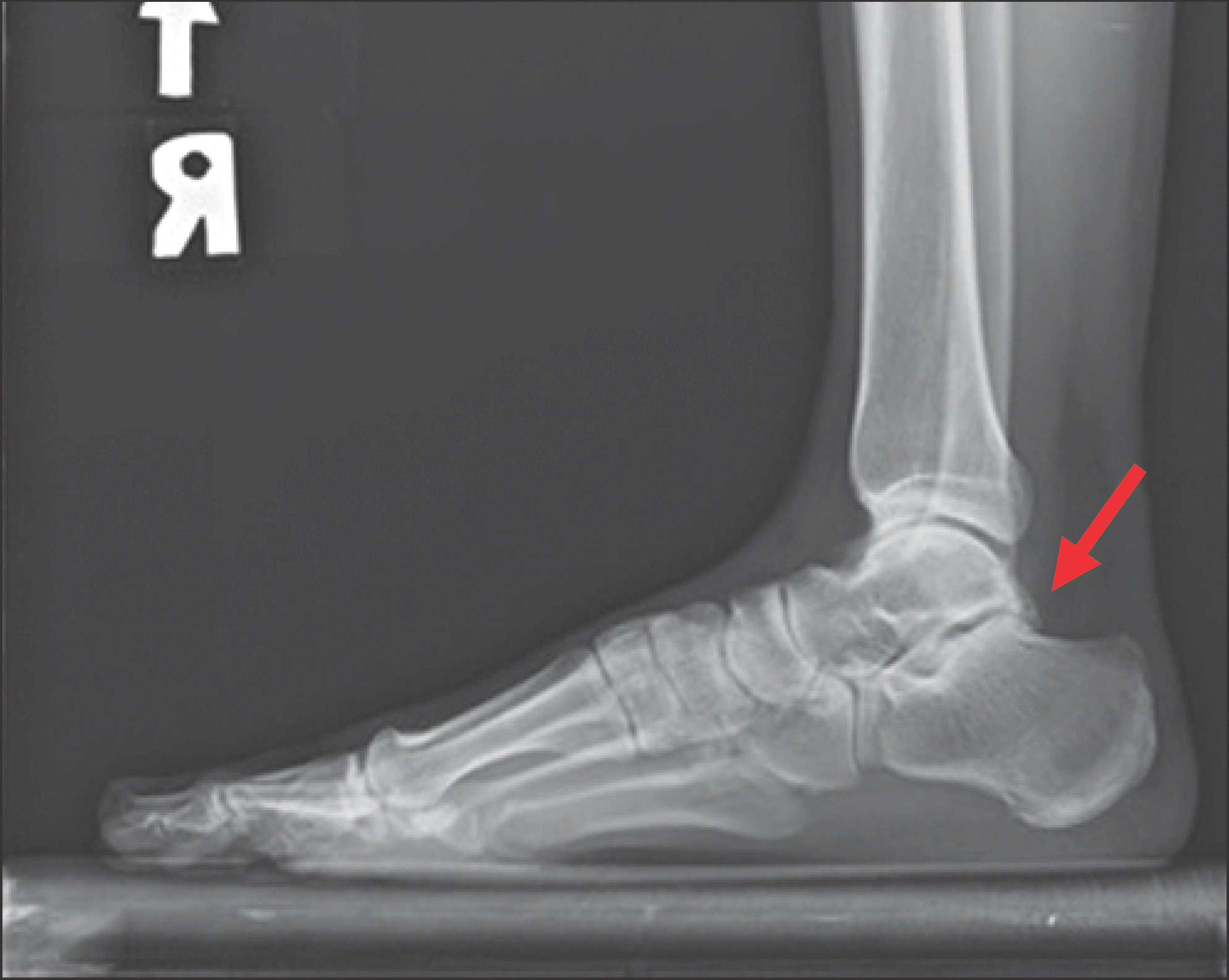
Figure 2.
Magnetic resonance images showing prominent osteophyte at posterior aspect of the talus at saggital view (A; arrow) and at axial view (B; arrow).
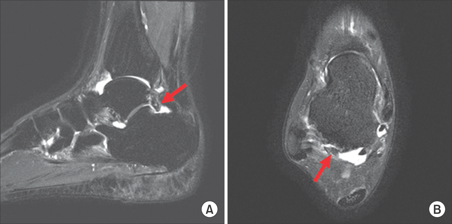
Figure 3.
Note the posteromedial cannula inserted for safety (A; arrow), and the cannula can be used for retracting and protecting the surrounding soft tissue, including the neurovascular bundle (B). v.: vein, FHL: flexor hallucis longus tendon, Med.: medial, Lat.: lateral, n.: nerve, Post.: posterior, a.: artery.
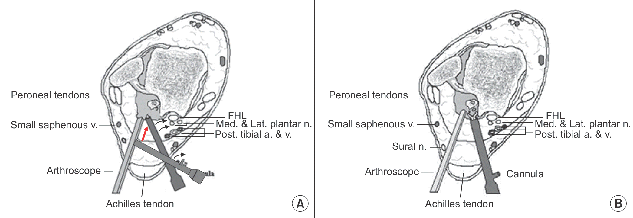
Figure 4.
The osteophyte and the loose body were visualized (A) and removed (B) through the posteromedial portal with a protection cannula.
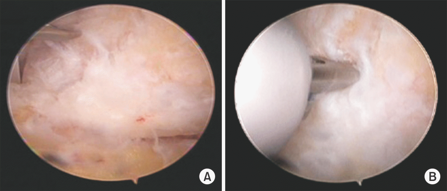
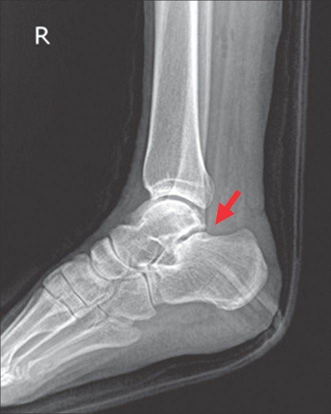
Table 1.
Preoperative Demographic Data in Both Groups
| Group A (n=25) | Group B (n=27) | p-value | |
|---|---|---|---|
| Age (yr) | 23.8±10.5 (15∼45) | 33.8±14.4 (17∼56) | 0.0502* |
| Sex (M:F) | 14:11 | 16:11 | 0.8139† |
| Baseline AOFAS score | 61±9 | 60±9 | 0.3435* |
| Follow-up period (mo) | 38.5±19.3 (24∼84) | 27.7±5.9 (24∼42) | 0.0283* |
Values are presented as mean±standard deviation (range), number only, or mean±standard deviation.
Group A was treated according to van Dijk et al.’s standard two-portal method,6) and group B was treated by a modification of the same method, using a protection cannula.
M: male, F: female, AOFAS: American Orthopaedic Foot and Ankle Society. *Mann-Whitney U-test.
Table 2.
The Comparison of the AOFAS Score between the Preoperative and Postoperative Periods in Each Group
| Preoperative period | Postoperative period | p-value | |
|---|---|---|---|
| AOFAS in group A (n=25) | 61±9 | 96±5 | <0.0000* |
| AOFAS in group B (n=27) | 60±9 | 94±5 | <0.0000* |
Values are presented as mean±standard deviation.
Group A was treated according to van Dijk et al.’s standard two-portal 6) method,6) and group B was treated by a modification of the same method, using a protection cannula.
AOFAS: American Orthopaedic Foot and Ankle Society. *Wilcoxon signed rank test.
Table 3.
The Time to Return to Activity in Both Groups
| Group A (n=25) | Group B (n=27) | p-value |
|---|---|---|
| Time (mo) 10.9±2.1 (8.3∼15.3) | 11.3±3.3 (7.5∼18.5) | ) 0.8397* |
Table 4.
Postoperative Complications in Both Groups
 XML Download