

 PDF
PDF ePub
ePub Citation
 Print
Print


Abstract
Dynamic conformal arc therapy (DCAT) and flattening-filter-free (FFF) beams are commonly adopted for efficient conformal dose delivery in stereotactic body radiation therapy (SBRT). Off-axis geometry (OAG) may be necessary to obtain full gantry rotation without collision, which has been shown to be beneficial for peripheral targets using flattened beams. In this study dose distributions in OAG using FFF were evaluated and the effect of mechanical rotation induced uncertainty was investigated. For the lateral target, OAG evaluation, sphere targets (2, 4, and 6 cm diameter) were placed at three locations (central axis, 3 cm off-axis, and 6 cm off-axis) in a representative patient CT set. For each target, DCAT plans under the same objective were obtained for 6X, 6FFF, 10X, and 10FFF. The parameters used to evaluate the quality of the plans were homogeneity index (HI), conformality indices (CI), and beam on time (BOT). Next, the mechanical rotation induced uncertainty was evaluated using five SBRT patient plans that were randomly selected from a group of patients with laterally located tumors. For each of the five cases, a plan was generated using OAG and CAG with the same prescription and coverage. Each was replanned to account for one degree collimator/couch rotation errors during delivery. Prescription isodose coverage, CI, and lung dose were evaluated. HI and CI values for the lateral target, OAG evaluation were similar for flattened and unflattened beams; however, 6FFF provided slightly better values than 10FFF in OAG. For all plans the HI and CI were acceptable with the maximum difference between flattened and unflattend beams being 0.1. FFF beams showed better conformality than flattened beams for low doses and small targets. Variation due to rotational error for isodose coverage, CI, and lung dose was generally smaller for CAG compared to OAG, with some of these comparisons reaching statistical significance. However, the variations in dose distributions for either treatment technique were small and may not be clinically significant. FFF beams showed acceptable dose distributions in OAG. Although 10FFF provides more dramatic BOT reduction, it generally provides less favorable dosimetric indices compared to 6FFF in OAG. Mechanical uncertainty in collimator and couch rotation had an increased effect for OAG compared to CAG; however, the variations in dose distributions for either treatment technique were minimal.
References
1. Timmerman R, Galvin J, Michalski J, Straube W, Ibbott G, Martin E, Abdulrahman R, Swann S, Fowler J, Choy H. Accreditation and quality assurance for Radiation Therapy Oncology Group: Multicenter clinical trials using Stereotactic Body Radiation Therapy in lung cancer. Acta Oncol. 2006; 45(7):779–786.

2. Takeda A, Kunieda E, Sanuki N, Ohashi T, Oku Y, Sudo Y, Iwashita H, Ooka Y, Aoki Y, Shigematsu N, Kubo A. Dose distribution analysis in stereotactic body radiotherapy using dynamic conformal multiple arc therapy. Int J Radiat Oncol Biol Phys. 2009; 74(2):363–369.
3. Cashmore J. The characterization of unflattened photon beams from a 6 MV linear accelerator. Phys Med Biol. 2008; 53(7):1933–1946.
4. Stathakis S, Esquivel C, Gutierrez A, Buckey CR, Papanikolaou N. Treatment planning and delivery of IMRT using 6 and 18MV photon beams without flattening filter. Appl Radiat Isot. 2009; 67(9):1629–1637.
5. Georg D, Knoos T, McClean B. Current status and future perspective of flattening filter free photon beams. Med Phys. 2011; 38(3):1280–1293.
6. Kragl G, Baier F, Lutz S, Albrich D, Dalaryd M, Kroupa B, Wiezorek T, Knoos T, Georg D. Flattening filter free beams in SBRT and IMRT: dosimetric assessment of peripheral doses. Z Med Phys. 2011; 21(2):91–101.
7. Thomas EM, Popple RA, Prendergast BM, Clark GM, Dobelbower MC, Fiveash JB. Effects of flattening filter-free and volumetricmodulated arc therapy delivery on treatment efficiency. J Appl Clin Med Phys. 2013; 14(6):4328.
8. Kretschmer M, Sabatino M, Blechschmidt A, Heyden S, Grunberg B, Wurschmidt F. The impact of flattening-filter-free beam technology on 3D conformal RT. Radiat Oncol. 2013; 8:133.
9. Ross CC, Kim JJ, Chen ZJ, Grew DJ, Chang BW, Decker RH. A novel modified dynamic conformal arc technique for treatment of peripheral lung tumors using stereotactic body radiation therapy. Pract Radiat Oncol. 2011; 1(2):126–134.
10. Shi C, Tazi A, Fang DX, Iannuzzi C. Implementation and evaluation of modified dynamic conformal arc (MDCA) technique for lung SBRT patients following RTOG protocols. Med Dosim. 2013; 38(3):287–290.
11. Kragl G, af Wetterstedt S, Knausl B, Lind M, McCavana P, Knoos T, McClean B, Georg D. Dosimetric characteristics of 6 and 10MV unflattened photon beams. Radiother Oncol. 2009; 93(1):141–146.
12. Ponisch F, Titt U, Vassiliev ON, Kry SF, Mohan R. Properties of unflattened photon beams shaped by a multileaf collimator. Med Phys. 2006; 33(6):1738–1746.
13. Yarahmadi M, Allahverdi M, Nedaie HA, Asnaashari K, Vaezzadeh SA, Sauer OA. Improvement of the penumbra for small radiosurgical fields using flattening filter free low megavoltage beams. Z Med Phys. 2013; 23(4):291–299.
14. Kim S. UF imageguided stereotactic body radiation therapy (IGSBRT) program for lung treatment using Elekta Synergy. In. IFMBE Proceedings. Edited by. Magjarevic R, editor. Springer;2007.
15. Olivier K, Chung H, Jin H, Yang H, Kim S. IntraFraction Patient Displacement During Image Guided Stereotactic Body Radiation Therapy (IGSBRT) for Lung Treatment. Int J Radiat Oncol Biol Phys. 2006; 66(3):): S489.
16. Khoo VS, Oldham M, Adams EJ, Bedford JL, Webb S, Brada M. Comparison of intensitymodulated tomotherapy with stereotactically guided conformal radiotherapy for brain tumors. Int J Radiat Oncol Biol Phys. 1999; 45(2):415–425.
17. Murphy MJ, Chang S, Gibbs I, Le QT, Martin D, Kim D. Image-guided radiosurgery in the treatment of spinal metastases. Neurosurg Focus. 2001; 11(6):): e6.
18. Josipovic M, Persson GF, Logadottir A, Smulders B, Westmann G, Bangsgaard JP. Translational and rotational intra-and interfractional errors in patient and target position during a short course of frameless stereotactic body radiotherapy. Acta Oncol. 2012; 51(5):610–617.
19. ACR-ASTRO practice guideline for the performance of stereotactic body radiation therapy. Practice Guidelines and Technical Standards. Reston, VA: American College of Radiology;2009.
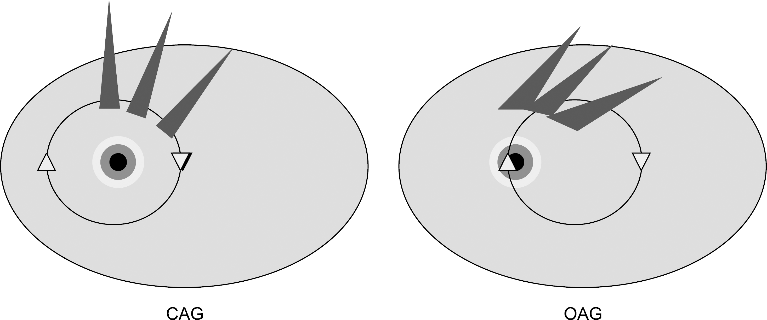
Fig. 2.
Lateral target, off-axis evaluation. Sphere targets of three sizes (2, 4, and 6 cm diameter) were placed at three locations (center of the patient, 3 cm lateral, and 6 cm lateral) for a total of 9 targets.
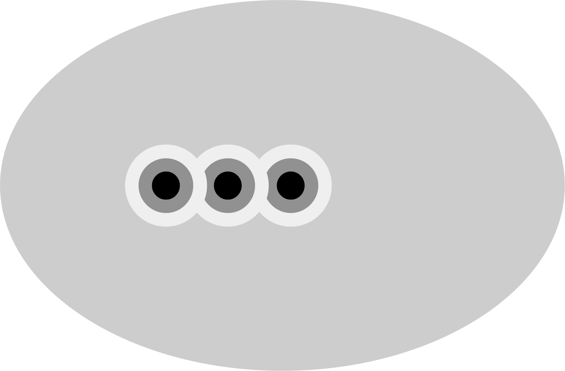
Fig. 3.
Mechanical rotation induced dosimetric uncertainty evaluation. Representative axial slice with OAG shown on the left and CAG on the right for each case.

Fig. 4.
Prescription Isodose Surface Coverage for off-axis geometry (OAG) and central axis geometry (CAG) for the original plan, one degree collimator rotation, one degree couch rotation, and one degree of both collimator and couch rotations. Points and lines for the same patient are shown in the same color.
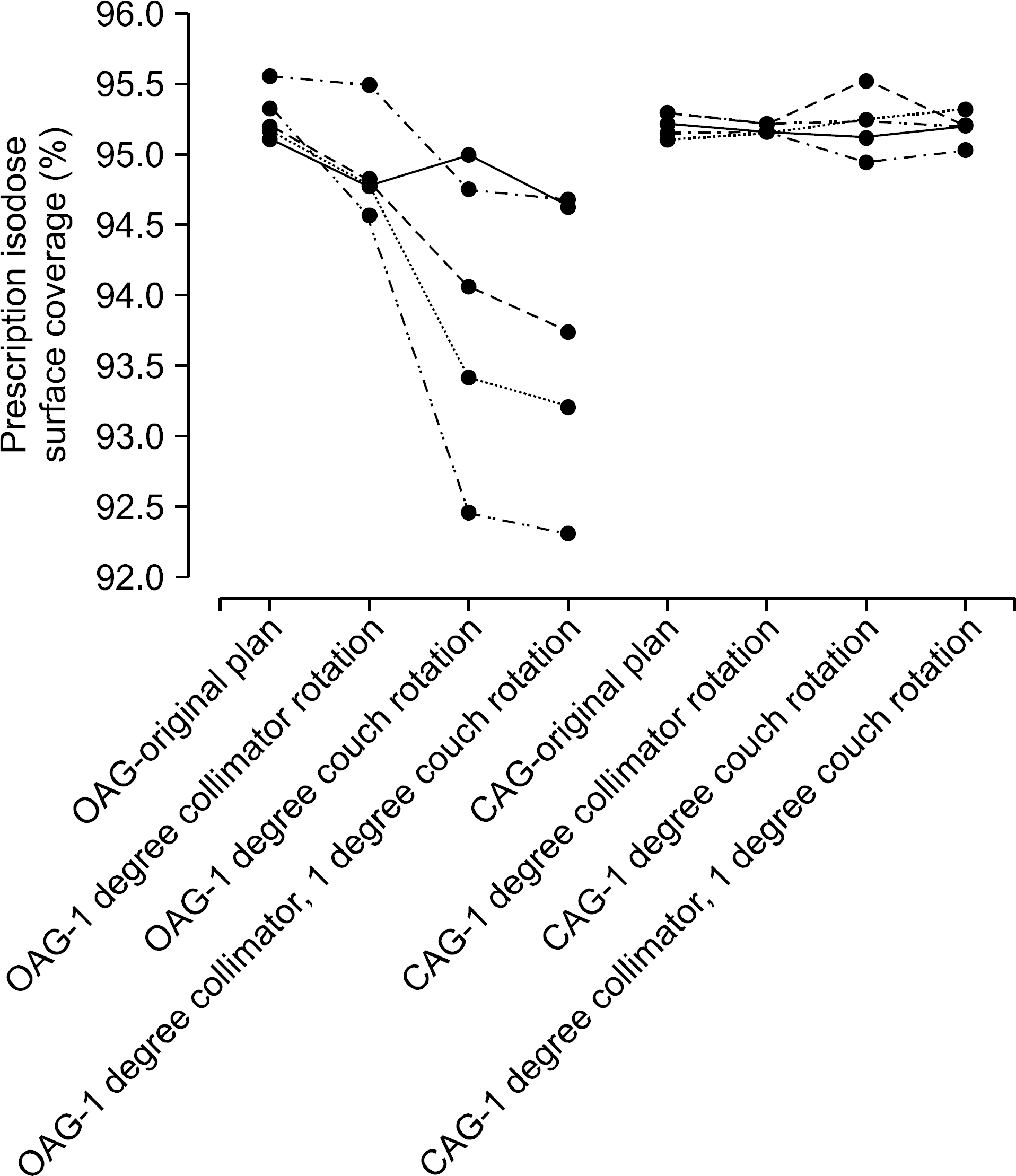
Fig. 5.
CI-100 for off-axis geometry (OAG) and central axis geometry (CAG) for the original plan, one degree collimator rotation, one degree couch rotation, and one degree of both collimator and couch rotations. Points and lines for the same patient are shown in the same color.
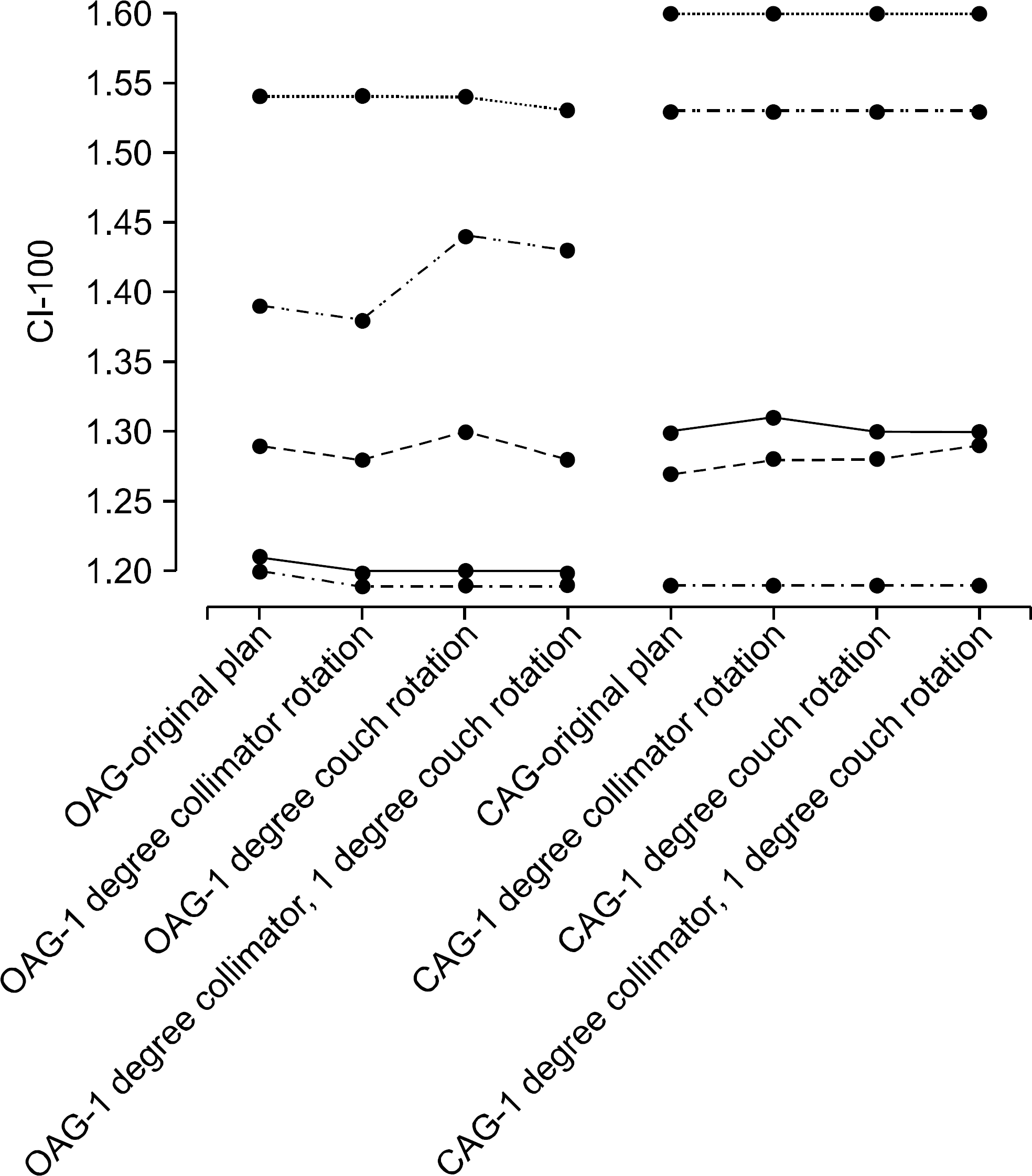
Fig. 6.
CI-50 for off-axis geometry (OAG) and central axis geometry (CAG) for the original plan, one degree collimator rotation, one degree couch rotation, and one degree of both collimator and couch rotations. Points and lines for the same patient are shown in the same color.
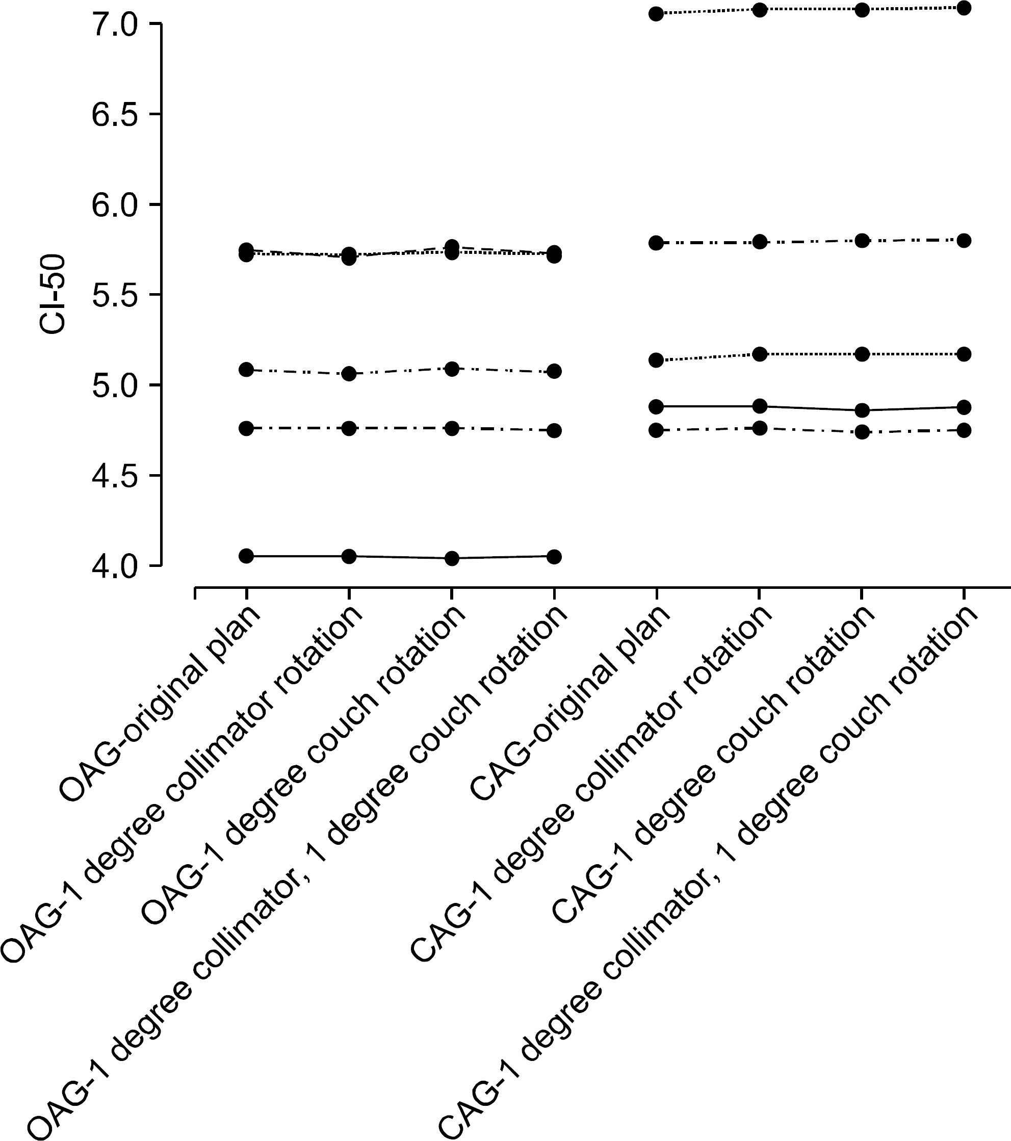
Fig. 7.
Lung V20 for off-axis geometry (OAG) and central axis geometry (CAG) for the original plan, one degree collimator rotation, one degree couch rotation, and one degree of both collimator and couch rotations. Points and lines for the same patient are shown in the same color.
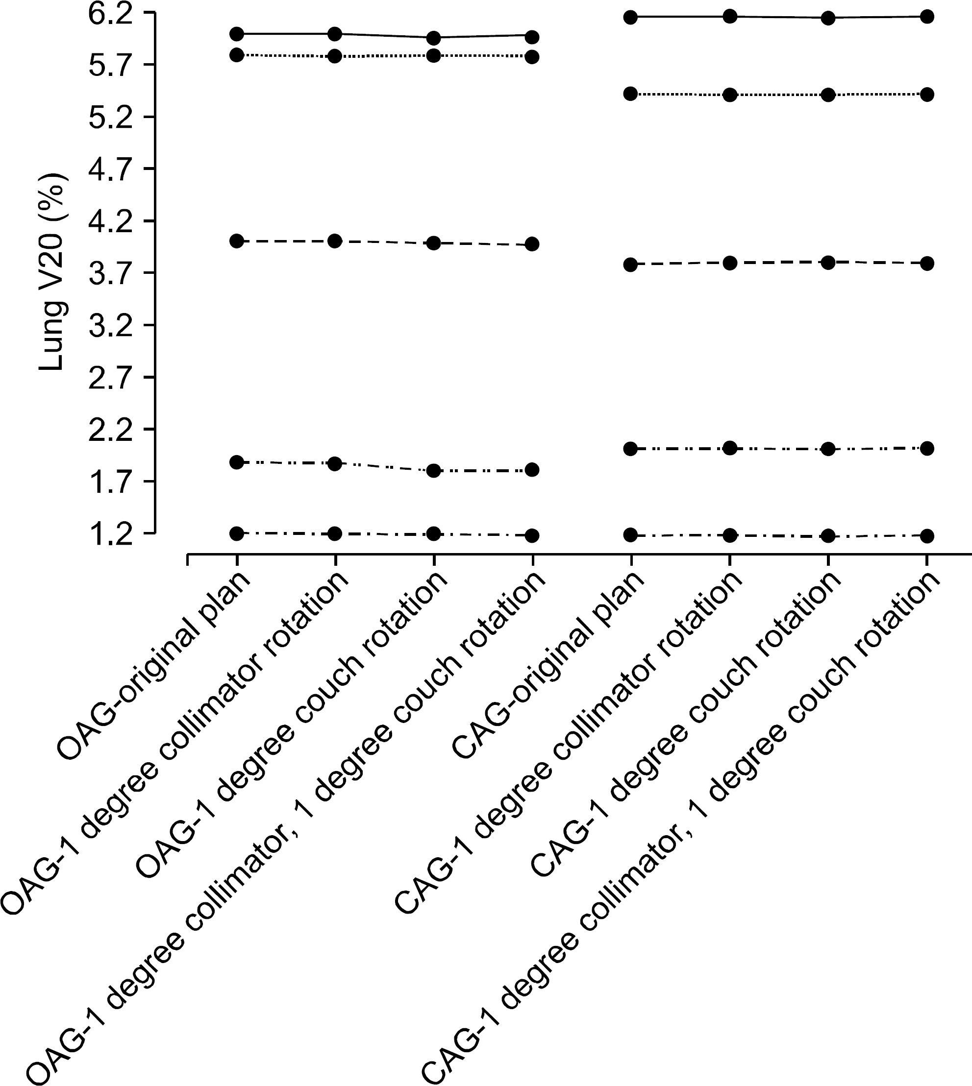
Table 1a.
Lateral target, off-axis geometry evaluation results for 6 MV.
Table 1b.
Lateral target, off-axis geometry evaluation results for 10 MV.
Table 1b.
Comparisons of prescription isodose surface coverage, conformality indices, and lung V20 between OAG and CAG.
 XML Download