

 PDF
PDF ePub
ePub Citation
 Print
Print


Abstract
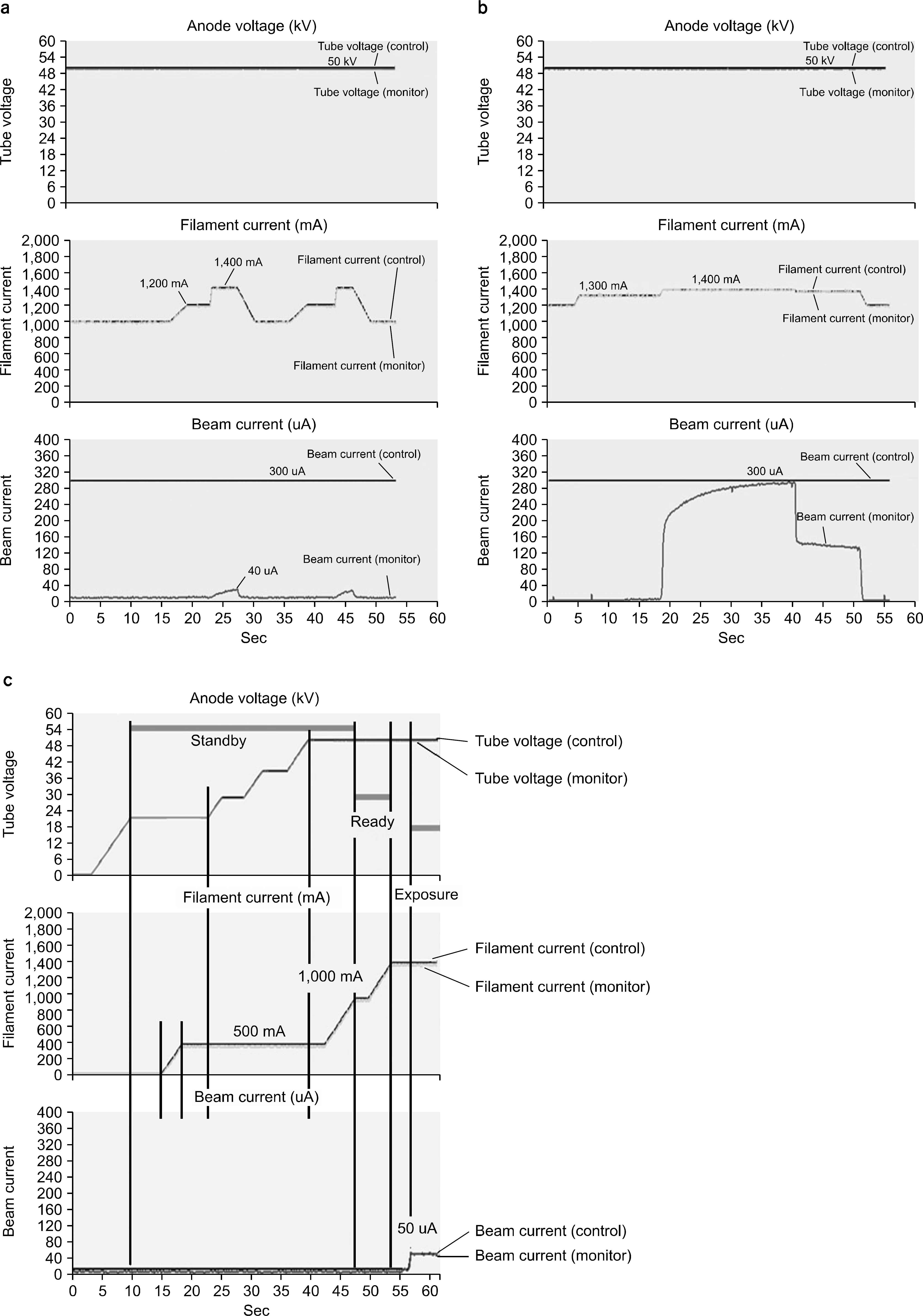
The purpose of this study is to develope a control system for a small X-ray tube generator and investigate control methods for the X-ray generator. The small X-ray tube was developed for electronic brachytherapy, and thus, the new control method should be investigated, if the small X-ray tube is used for the imaging system. The Axxent S700 X-ray tube and the XF060NZZ485 high voltage generator were used to compose a X-ray imaging system and control board was developed by using AT90CAN128 MCU. The two control methods were investigated after tube voltage was set to 50 kV, one was filament current control method and the other was beam current control method. The former was subdivided into two methods according to the filament heating time, the 5 and the 10 seconds respectively. In the filament current method, the beam current did not rise up to the desired value, if the filament current had not been maintained for at least 10 seconds. The onset filament currents to generate beam current were varied from 1,300 to 1,350 mA and over 5 seconds were needed in order to reach the desired tube current value after beam current was generated. However, in the tube current control method, the beam current reached to the desired value without any time delay with the filament current of 1,500 mA. In this study, we found that the beam current control method was appropriate for the use of small X-ray tube developed for brachytherapy in the X-ray imaging system.
References
1. Wenzel A, Hintze H, Mikkelsen L, et al. Radiographic detection of occlusal caries in noncavitated teeth: a comparison of conventional film radiographs, digitized film radiographs and radiography. Oral Surg Oral Med Oral Pathol. 72(5):621–626. 1991.
2. Shearer AC, Homer K, Wilson NH. Radiovisiography for imaging root canals: an in vitro comparison with conventional radiography. Quint Int. 21(10):789–794. 1990.
3. Furkart AJ, Dove SB, McDavid WD, et al. Direct digital radiography for detection of periodontal bone lesions. Oral Surg Oral Med Oral Pathol. 74(5):652–660. 1992.
4. Cho SH, Kim DY, Baek KW. Introduction of Dental X-rayImaging with New Concept –intra Oral x-ray Tube. J Inst Electron Engineer Korea. 47:(. (SP3):):. 70–80. 2010.
5. Cho SH, Kim SY, An SH. Feasibility study of insertable miniature x-ray source for dental imaging. J Korean Soc Radiol. 6(1):39–45. 2012.

6. Dickler A. Xoft Axxent electronic brachytherapy: a new device for delivering brachytherapy to the breast. Nat Clin Pract Oncol. 6(3):138–142. 2009.
7. Dickler A, Kirk MC, Sief N, et al. A dosimetric comparison of MammoSite high-dose-rate brachytherapy and Xoft Axxent electronic brachytherapy. Brachytherapy. 6(2):164–168. 2007.
8. Dickler A, Dowlatshahi K. Xoft Axxent electronic brachytherapy. Expert Rev Med Devices. 6(1):27–31. 2009.
Fig. 1.
Photographies of intra-oral X-ray tube: (a) X-ray source, (b) X-ray tube module, and (c) HV generator & cable.
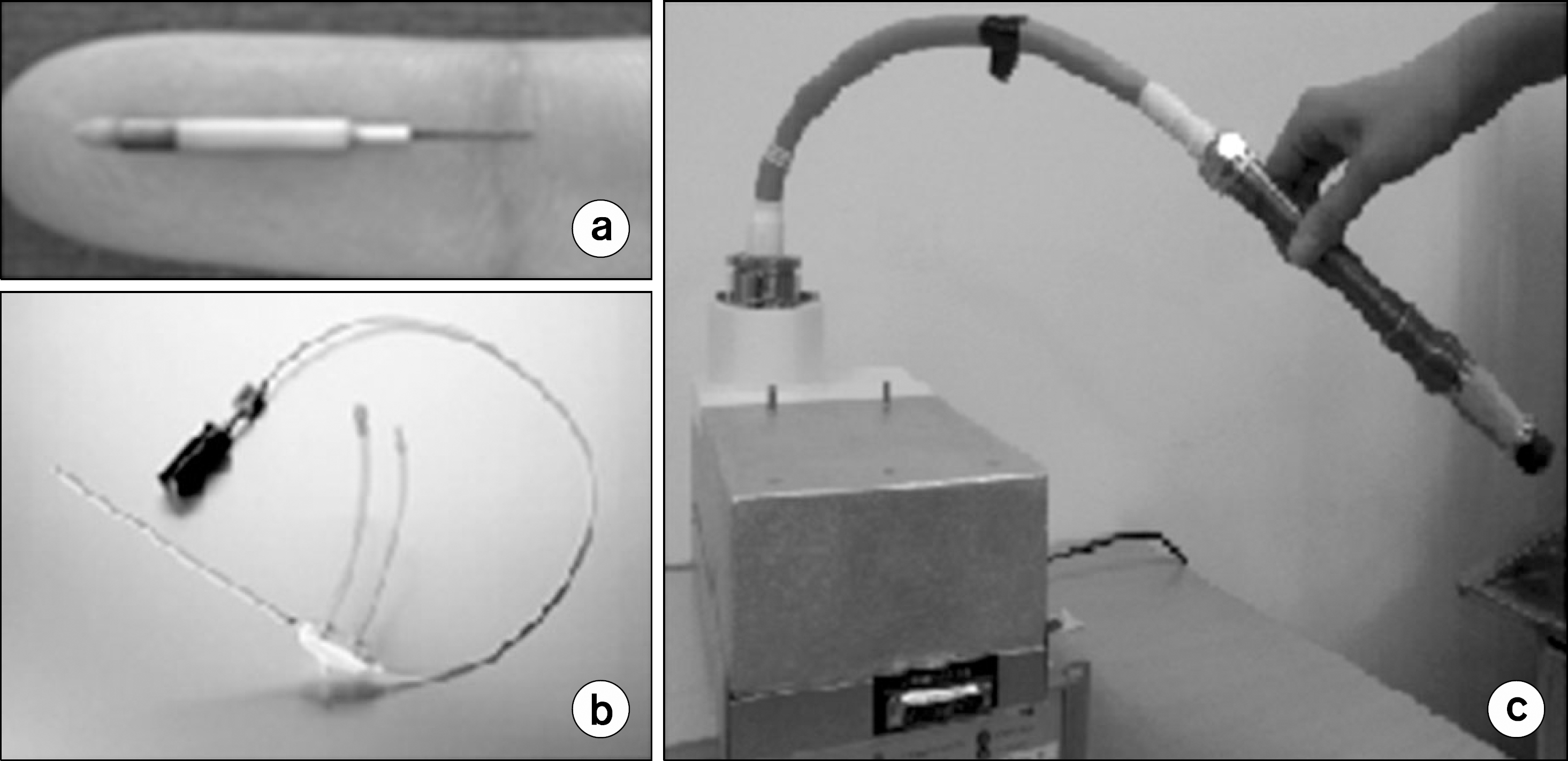
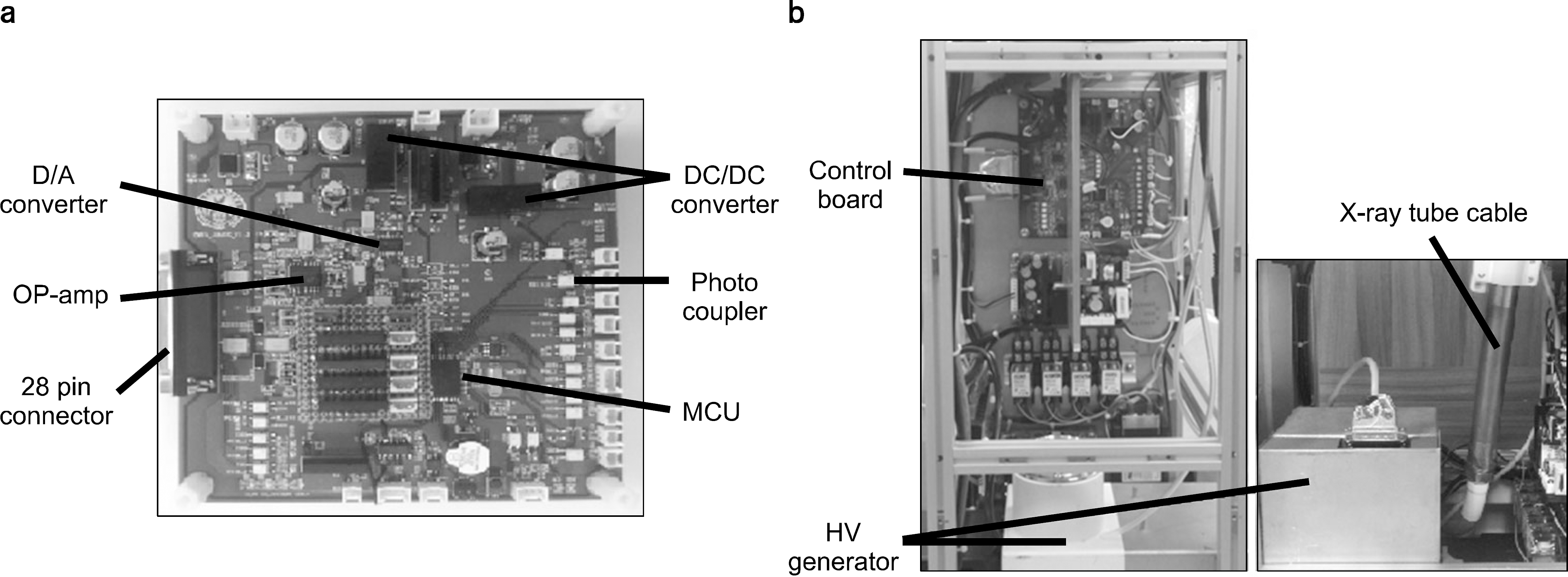
Fig. 2.
Photographies of HV generator control System: (a) Control board and (b) HV generator system.
 XML Download