

 PDF
PDF ePub
ePub Citation
 Print
Print


Abstract
Previous studies about effect of respiratory motion on diagnostic imaging and radiation therapy have been performed by monitoring external motions but these can not reflect internal organ motion well. The aim of this study was to develope the artificial pulmonary nodule able to perform non-invasive implantation to dogs in the thorax and to evaluate applicability of the model to respiratory motion studies on PET image acquisition and radiation delivery by phantom studies. Artificial pulmonary nodule was developed on the basis of 8 Fr disposable gastric feeding tube. Four anesthetized dogs underwent implantation of the models via trachea and implanted locations of the models were confirmed by fluoroscopic images. Artificial pulmonary nodule models for PET injected18F-FDG and mounted on the respiratory motion phantom. PET images of those acquired under static, 10-rpm- and 15-rpm-longitudinal round motion status. Artificial pulmonary nodule models for radiation delivery inserted glass dosemeter and mounted on the respiratory motion phantom. Radiation delivery was performed at 1 Gy under static, 10-rpm- and 15-rpm-longitudinal round motion status. Fluoroscpic images showed that all models implanted in the proximal caudal bronchiole and location of models changed as respiratory cycle. Artificial pulmonary nodule model showed motion artifact as respiratory motion on PET images. SNR of respiratory gated images was 7.21. which was decreased when compared with that of reference images 10.15. However, counts of respiratory images on profiles showed similar pattern with those of reference images when compared with those of static images, and it is assured that reconstruction of images using by respiratory gating improved image quality. Delivery dose to glass dosemeter inserted in the models were same under static and 10-rpm-longitudinal motion status with 0.91 Gy, but dose delivered under 15-rpm-longitudinal motion status was decreased with 0.90 Gy. Mild decrease of delivered radiation dose confirmed by electrometer. The model implanted in the proximal caudal bronchiole with high feasibility and reflected pulmonary internal motion on fluoroscopic images. Motion artifact could show on PET images and respiratory motion resulted in mild blurring during radiation delivery. So, the artificial pulmonary nodule model will be useful tools for study about evaluation of motion on diagnostic imaging and radiation therapy using laboratory animals.
References
1. Phelps ME. Positron emission tomography provides molecular imaging of biological processes. Proc Natl Acad Sci. 97(16):9226–9233. 2000.

2. Bryan PJ, Custar S, Haaga JR, Balsara V. Respiratory movement of the pancreas: an ultrasonic study. J Ultrasound Med. 3(7):317–320. 1984.
3. Ross CS, Hussey DH, Penington EC, et al. Analysis of movement of intrathoracic neoplasms using ultrafast computed tomography. Int J Radiat Oncol Biol Phys. 18(3):671–677. 1990.
4. Davies SC, Hill AL, Holmes RB, Halliwell M, Jackson PC. Ultrasound quantitation of respiratory organ motion in the upper abdomen. Br J Radiol. 67(803):1096–1102. 1994.
5. Nehmeh SA, Erdi YE. Respiratory motion in positron emission tomography/computed tomography: Review. Semin Nucl Med. 38:167–176. 2008.
6. Chun SY, Reese TG, Ouyang JS, et al. MRI-Based nonrigid motion correction in simultaneous PET/MRI. J Nucl Med. 53(8):1284–1291. 2012.
7. Osman MM, Cohade C, Nakamoto Y, Wahl RL. Respiratory motion artifacts on PET emission images obtained using CT attenuation correction on PET-CT. Eur J Med Mol Imaging. 30(4):603–606. 2003.
8. Catana C, Benner T, van der Kouwe A, et al. MRI-assisted PET motion correction for neurologic studies in an integrated MR-PET scanner. J Nucl Med. 52(1):154–161. 2011.
9. Zhang Q, Pevsner A, Hertanto A, et al. A pateint-specific respiratory model of anatomical motion for radiation treatment planning. Med Phys. 34(12):4772–4781. 2007.
10. Caldwell CB, Mah K, Skinner M, Danjoux CE. Can PET provide the 3D extent of tumor motion for individualized internal target volumes? A phantom study of the limitations of CT and the promise of PET. Int J Radiation Oncology Biol Phys. 55(5):1381–1393. 2003.
11. Low DA, Parikh PJ, Lu W, et al. Novel breathing motion model for radiotherapy. Int J Radiation Oncology Biol Phys. 63(3):921–929. 2005.
12. Yu JW, Woo SK, Lee YJ, et al. Estimation of internal motion for quantitative improvement of lung tumor in small animal. Korean J Med Phys. 22(3):140–147. 2011.
13. Ionascu D, Jiang SB, Nishioka S, Hiroki S, Berbeco RI. Internal-external correlation investigations of respiratory induced motion of lungs. Med Phys. 34(10):3893–3903. 2007.
14. ASAHI Glass Cor. 2004. Explanation Material of RPL Glass Dosimeter: Small Element System. Tokyo, Japan.
15. Eom K, Seoung Y, Park H, Choe N, Park J, Jang K. Radiologic and computed tomographic evaluation experimentally induced lung aspiration sites in dogs. J Vet Sci. 7(4):397–399. 2006.
16. Bortfeld T, Jiang SB, Rietzel E. Effects of motion on the total dose distribution. Semin Radiat Oncol. 14(1):41–51. 2004.
Fig. 1.
Artificial pulmonary nodule models for (a): PET and (b): radiation delivery; (a), the nodule part of the model for PET phantom study in which cotton (∗) was inserted; (b), the nodule part of the model for radiation delivery phantom study in which glass dosemeter (†) was inserted.
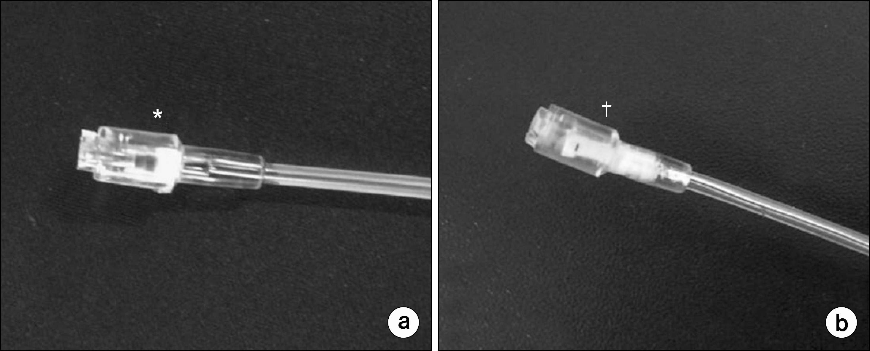
Fig. 2.
The set up for respiratory motion studies and represented fluoroscopic images. (a) the setup for respiratory motion study on the PET with the model mounted on the respiratory phantom; (b) the setup for respiratory motion study on the radiation delivery with the model mounted on the respiratory phantom.
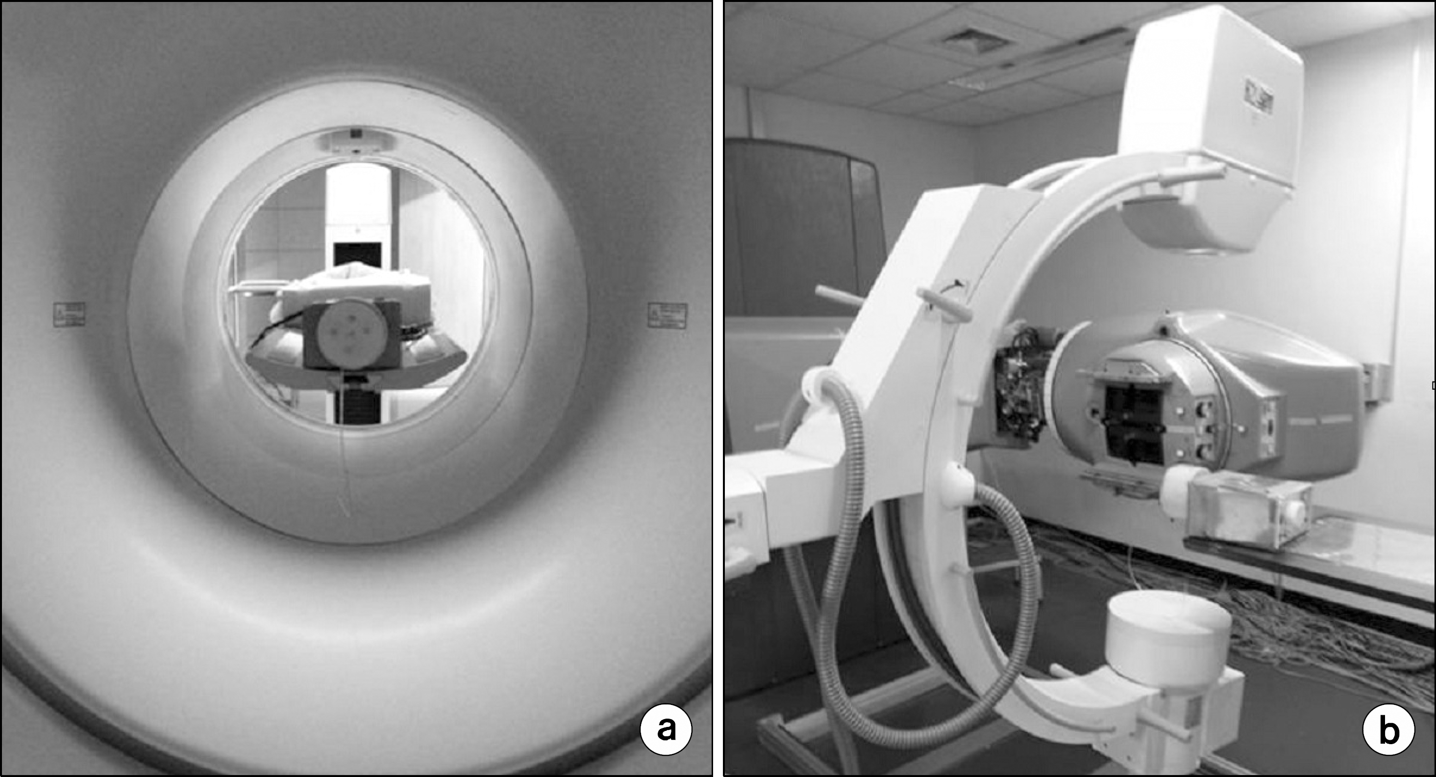
Fig. 3.
Fluoroscopic images of artificial pulmonary nodule models implantation in 4 dogs (from top to bottom). (a) fluoroscopic images of inserted artificial pulmonary nodule models (white star) in dogs at inspiration. (b) fluoroscopic images of inserted artificial pulmonary nodule models (white star) in dogs at expiration.

Fig. 4.
Acquired images of artificial pulmonary nodule model by PET. (a), reference images; (b), static images; (c), respiratory gated images; left, transaxial images; middle, coronal images; right, saggital images.
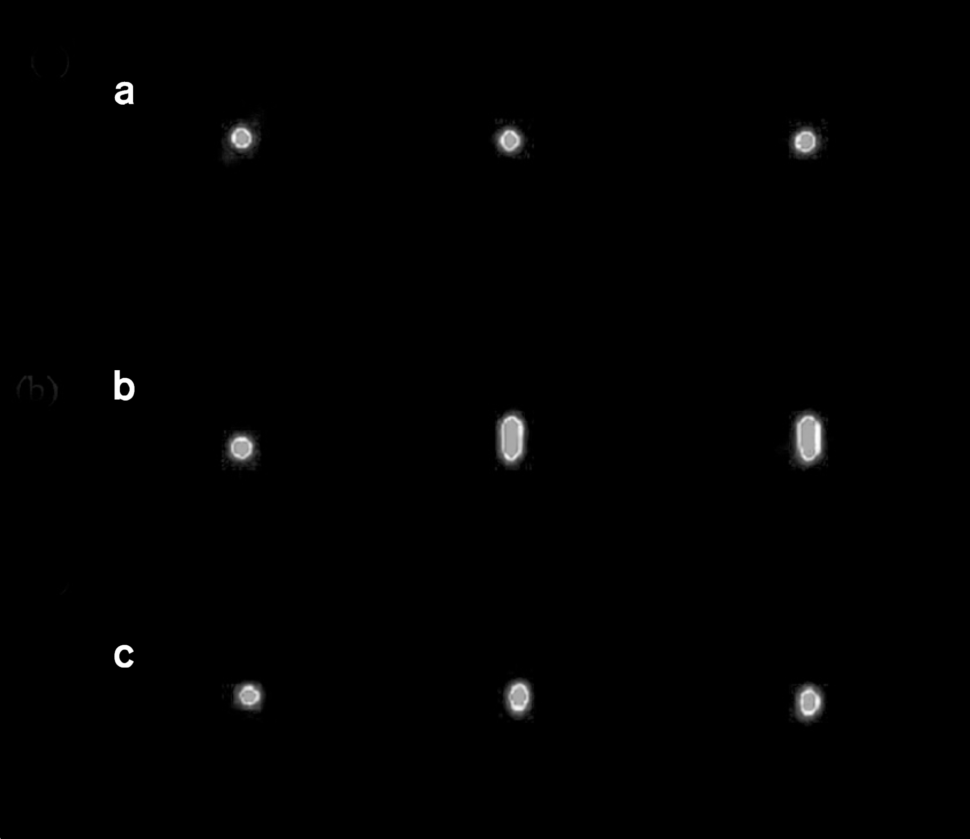
Fig. 5.
Quantitative analysis of motion in PET images. (a), estimated SNR to reference, static, gated images of artificial pulmonary nodule models at 15-rpm-longitudinal motion; (b), the count of vertical line profile drawn at the artificial pulmonary nodule model during motion at 15 rpm. The diamond line, the square line and the triangle line represent as data of reference, static image and gated image, respectively.
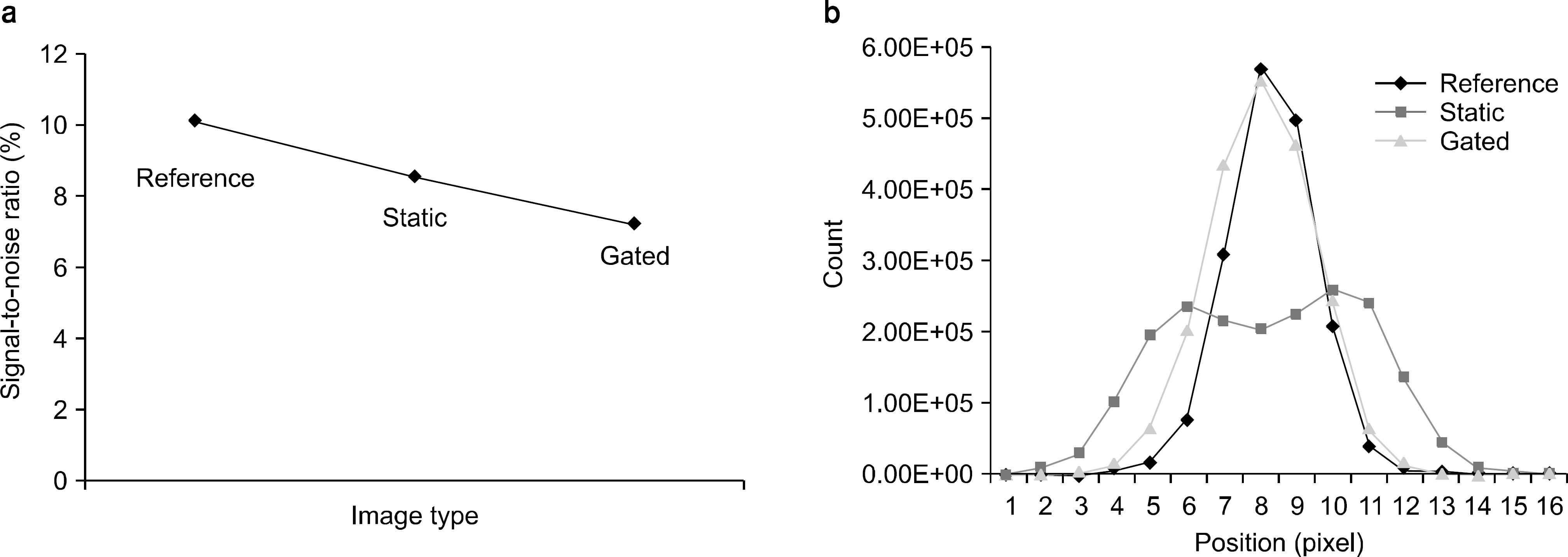
Fig. 6.
Relationship of SNR of respiratory gated PET images and delivered radiation dose as motion rate changes of phantom. The diamond and square represent as SNR and delivered radiation dose, respectively. The blue and red lines represent as drift curves of SNR and delivered radiation dose, respectively. Reference, motion status under rest; N-10, sine wave motion at 10 rpm; N-15, sine wave motion at 15 rpm; R-10, respiratory motion at 10 rpm; R-15, respiratory motion at 15 rpm.
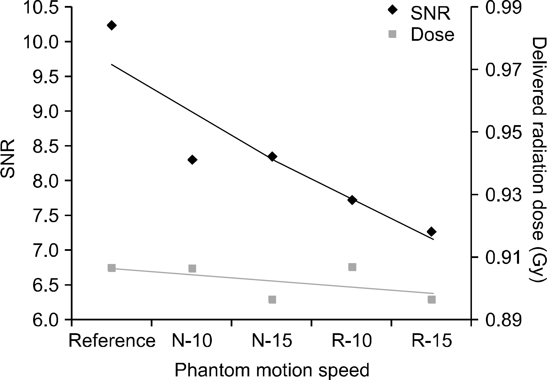
Table 1.
Delivered radiation dose to artificial pulmonary nodule models mounted respiratory phantom with static, 10-and 15-rpm-longitudinal round motion.
| Glass dosemeter | Ion chamber | |||
|---|---|---|---|---|
| Dose (Gy) | Difference (%) | Dose (nC) | Difference (%) | |
| Reference | 0.91 | – | 13.91 | – |
| 10 rpm | 0.91 | 0 | 13.91 | 0 |
| 15 rpm | 0.90 | 1.10 | 13.87 | 0.28 |
 XML Download