

 PDF
PDF ePub
ePub Citation
 Print
Print


Abstract
In the intracranial regions, an accurate delineation of the target volume has been difficult with only the CT data due to poor soft tissue contrast of CT images. Therefore, the magnetic resonance images (MRI) for the delineation of the target volumes were widely used. To calculate dose distributions with MRI-based RTP, the electron density (ED) mapping concept from the diagnostic CT images and the pseudo CT concept from the MRI were introduced. In this study, the look up table (LUT) from the fifteen patients’ diagnostic brain MRI images was created to verify the feasibility of MRI-based RTP. The dose distributions from the MRI-based calculations were compared to the original CT-based calculation. One MRI set has ED information from LUT (lMRI). Another set was generated with voxel values assigned with a homogeneous density of water (wMRI). A simple plan with a single anterior 6MV one portal was applied to the CT, lMRI, and wMRI. Depending on the patient's target geometry for the 3D conformal plan, 6MV photon beams and from two to five gantry portals were used. The differences of the dose distribution and DVH between the lMRI based and CT-based plan were smaller than the wMRI-based plan. The dose difference of wMRI vs. lMRI was measured as 91 cGy vs. 57 cGy at maximum dose, 74 cGt vs. 42 cGy at mean dose, and 94 cGy vs. 53 at minimum dose. The differences of maximum dose, minimum dose, and mean dose of the wMRI-based plan were lower than the lMRI-based plan, because the air cavity was not calculated in the wMRI-based plan. These results prove the feasibility of the lMRI-based planning for brain tumor radiation therapy.
REFERENCES
1. Jonsson JH, Karlsson MG, Karlsson M, Nyholm T. Treatment planning using MRI data: an analysis of the dose calculation accuracy for different treatment regions. Radiat Oncol. 5:62. 2010.

2. Prabhakar R, Julka PK, Ganesh T, Munshi A, Joshi RC, Rath G. Feasibility of using MRI alone for 3D radiation treatment planning in brain tumors. Jpn J Clin Oncol. 37(6):405–411. 2007.
3. Stanescu T, Hans-Sonke J, Stavrev P, Fallone BG. 3T MR-based treatment planning for radiotherapy of brain lesions. Radiat Oncol. 40(2):125–132. 2006.
4. Chen L, Price RA Jr, Wang L, et al. MRI-based treatment planning for radiotherapy: dosimetric verification for prostate IMRT. Int J Radiat Oncol Biol Phys. 60(2):636–647. 2004.
5. Wang C, Chao M, Lee L, Xing L. MRI-based treatment planning with electron density information mapped from CT. Technol Cancer Res Treat. 7(5):341–347. 2008.
6. Miga MI, Boettger T, Nyholm T, et al. Radiation therapy planning and simulation with magnetic resonance images. Medical Imaging. 6918:19181C. 2008.
Fig. 1.
The OARs are contoured on the magnetic resonance images (MRI) in order to define the gray scales; (a) The brain soft tissue, (b) Eyeball, (c) Nasal cavity, and (d) Bone.
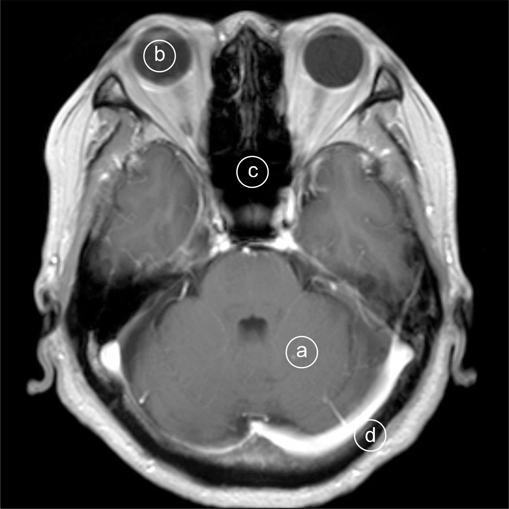
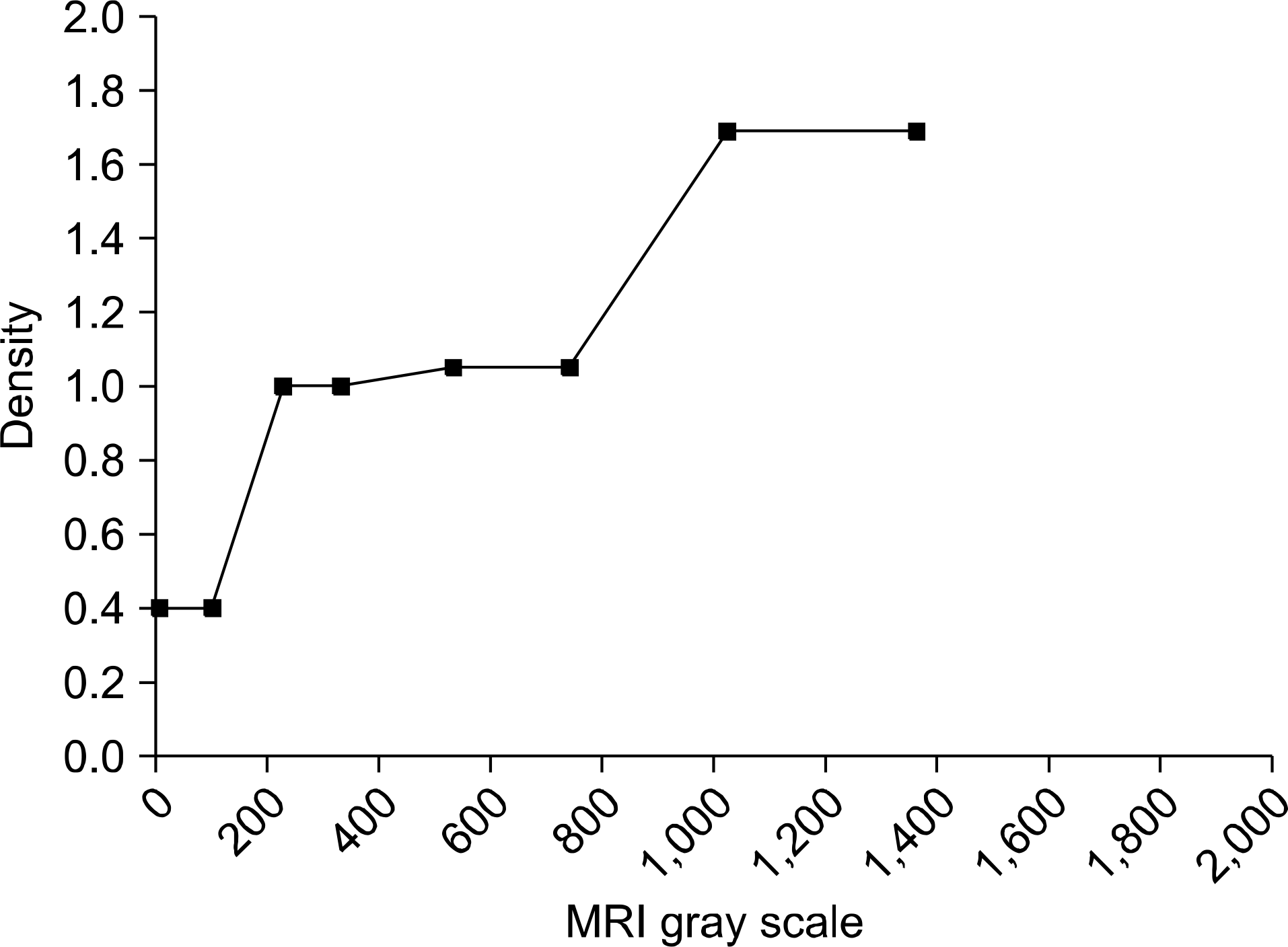
Fig. 3.
Dose distributions are shown on the CT image and MR images (a) CT-based isodose curve, (b) lMRI-based isodose curve, and (c) wMRI-based isodose curve.

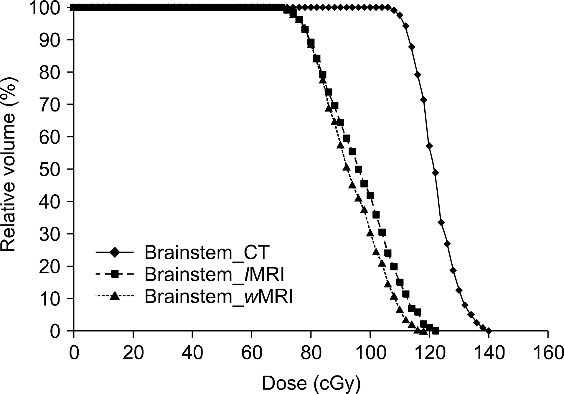
Fig. 5.
15 patients average maximum dose difference between wMRI and lMRI based plans. The reference dose were CT-based plans.
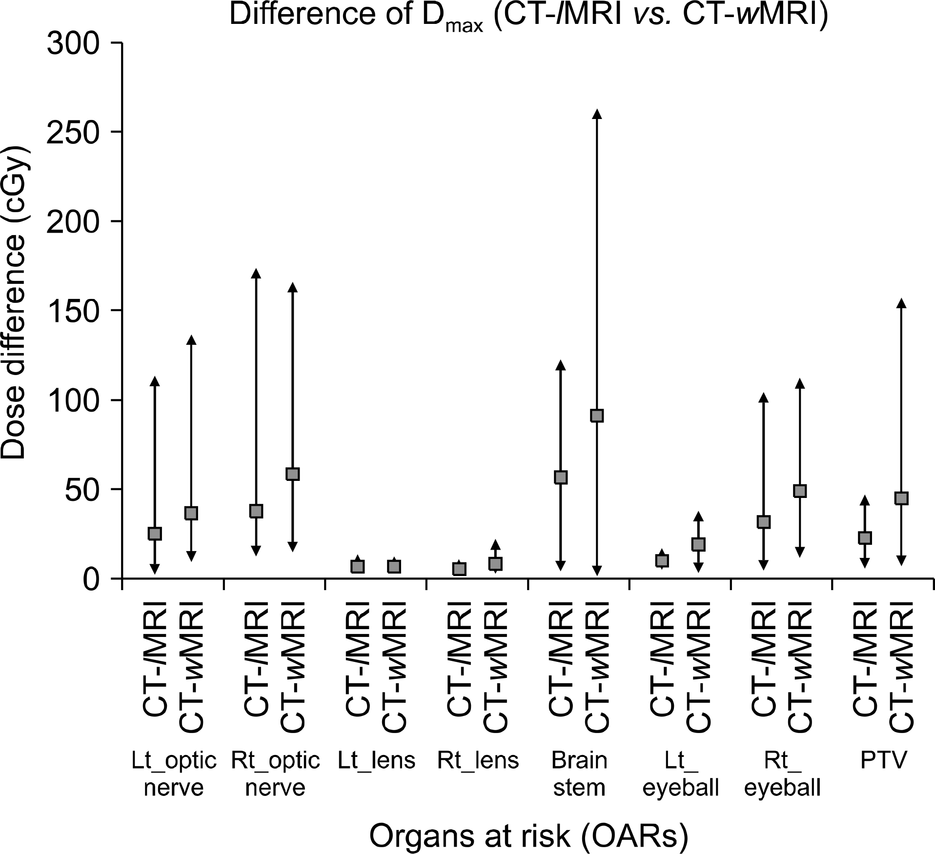
Fig. 6.
15 patients average mean dose difference between wMRI and lMRI based plans. The reference dose were CT-based plans.
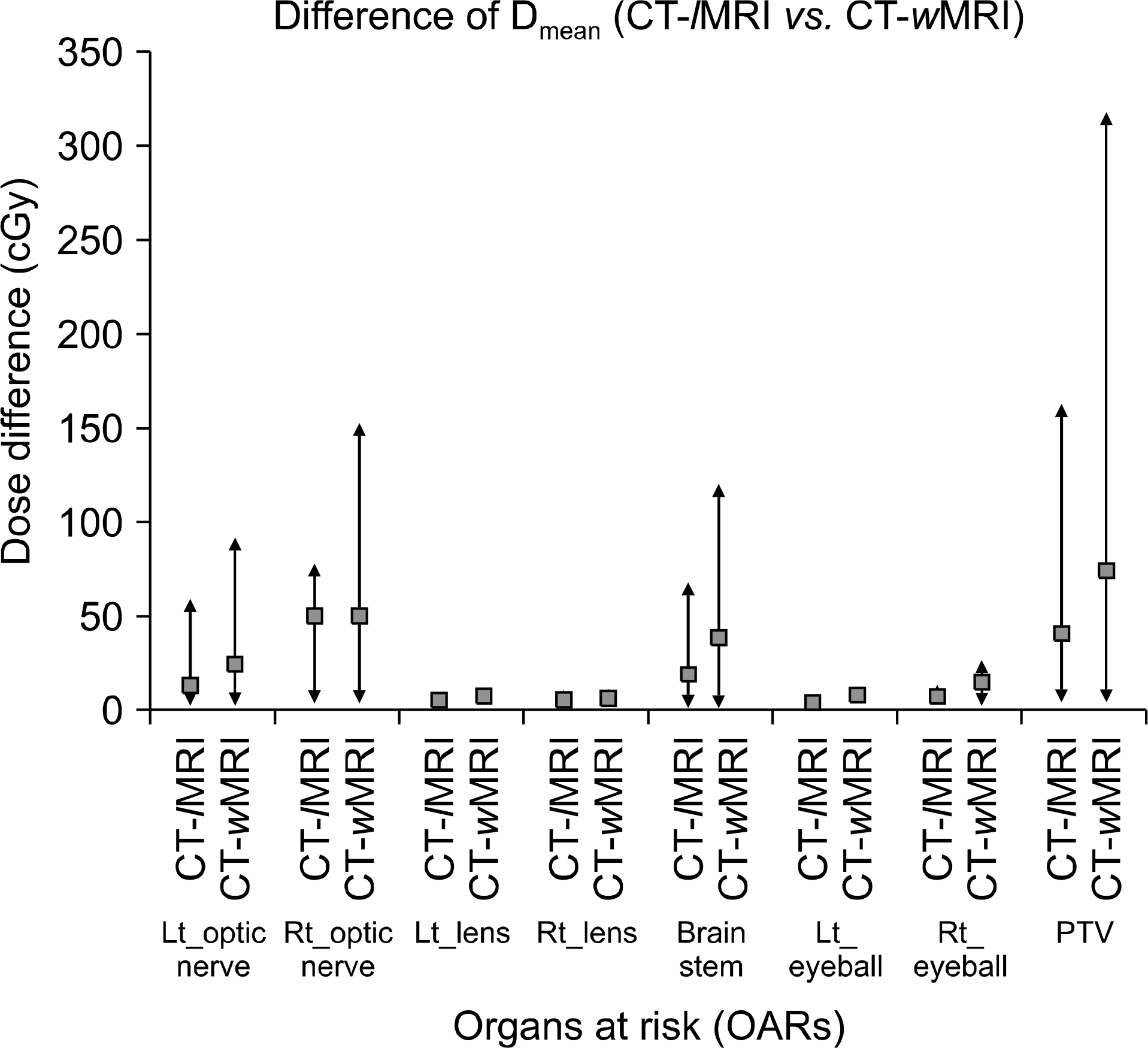
Fig. 7.
15 patients average minimum dose difference between wMRI and lMRI based plans. The reference dose were CT-based plans.
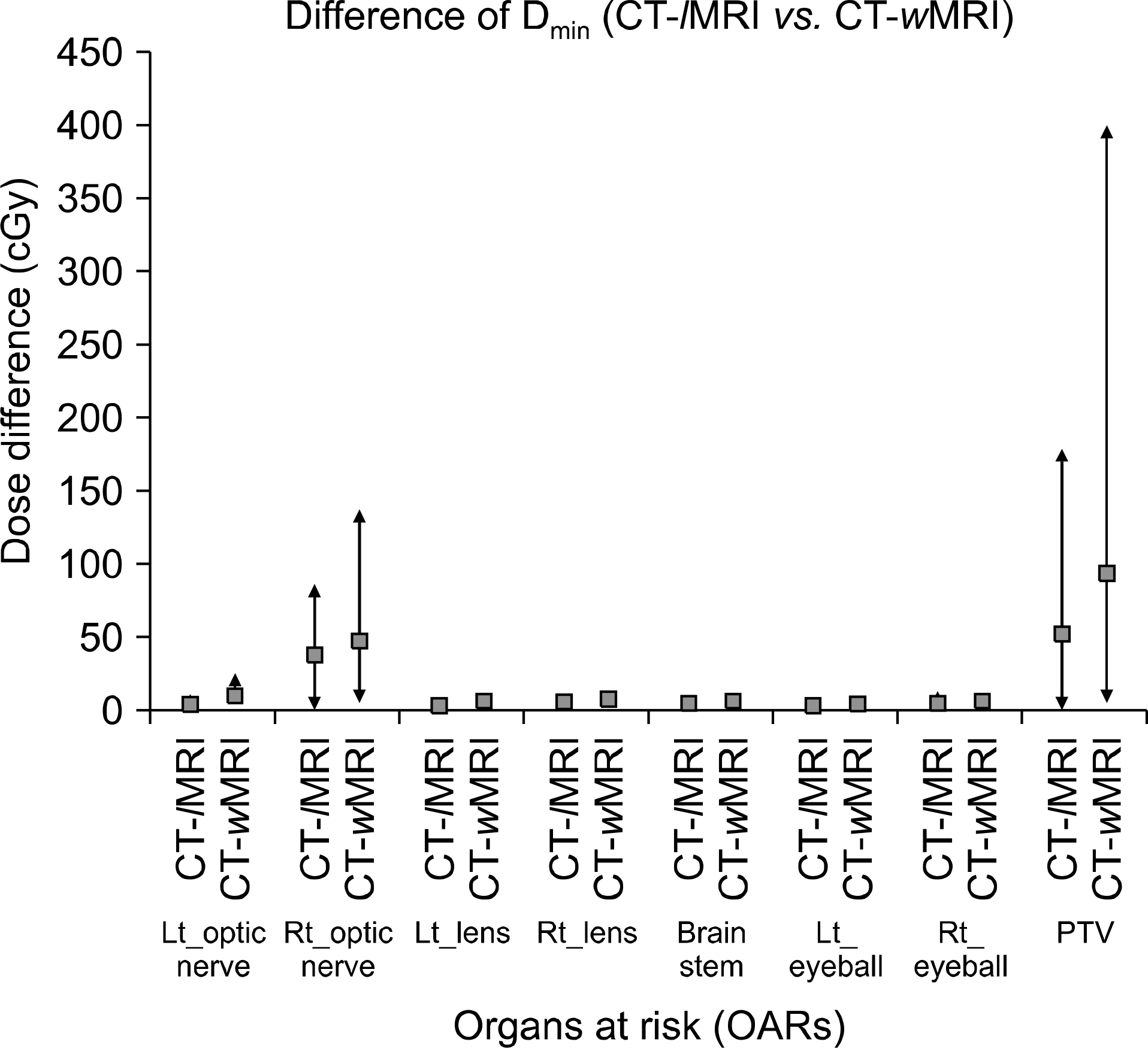
Table 1.
General patients’ information and patients’ gray scale data at OARs.
| Patients | Age | Gender | Diagnosis | Brain | Eyeball | Cavity | Bone |
|---|---|---|---|---|---|---|---|
| 1 | 57 | Male | Glioblastoma | 739 | 329 | 98 | 1,277 |
| 2 | 49 | Male | Glioblastoma | 701 | 283 | 21 | 1,362 |
| 3 | 51 | Male | Anaplastic astrocytoma | 747 | 306 | 54 | 1,261 |
| 4 | 63 | Female | Anaplastic oligdendroglioma | 778 | 328 | 15 | 1,414 |
| 5 | 38 | Female | Glioblastoma | 721 | 318 | 17 | 1,119 |
| 6 | 41 | Male | Glioblastoma | 616 | 307 | 9.9 | 1,217 |
| 7 | 71 | Female | Anaplastic oligdendroglioma | 624 | 294 | 65 | 1,223 |
| 8 | 38 | Female | Glioblastoma | 539 | 226 | 16 | 1,024 |
| 9 | 44 | Male | Glioblastoma | 694 | 305 | 19 | 1,178 |
| 10 | 50 | Male | Anaplastic oligdendroglioma | 719 | 307 | 39 | 1,152 |
| 11 | 51 | Female | Glioblastoma | 645 | 292 | 45 | 1,220 |
| 12 | 74 | Female | Glioblastoma | 637 | 275 | 14 | 1,182 |
| 13 | 81 | Female | Glioblastoma | 533 | 235 | 19 | 1,116 |
| 14 | 35 | Male | Anaplastic astrocytoma | 658 | 293 | 9.7 | 1,348 |
| 15 | 41 | Female | Anaplastic astrocytoma | 652 | 294 | 9.6 | 1,207 |
| Range | 35∼81 | 533∼739 | 226∼329 | 9.6∼98 | 1,024∼1,362 | ||
| Average | 52.3 | 666.9 | 292.8 | 30.1 | 1,220.0 | ||
| SD∗ | 14.2 | 71.7 | 29.4 | 25.5 | 102.3 |
 XML Download