

 PDF
PDF ePub
ePub Citation
 Print
Print


Abstract
Recent developments of image guided radiation therapy (IGRT), especially the On Board Imaging (OBI) system and the cone beam CT (CBCT), enable the radiation treatment more accurate and reliable. IGRT is widely used in the radiation therapy as a standard of care. Use of IGRT is even expected to increase in the near future. IGRT is only beneficial to patients when it is used with proper considerations of safety and appropriateness of the techniques. Institutional procedure should be developed based on the clinical need and the deep understanding of the system before applying the new technique to the clinic. Comprehensive QA program should be established before to the clinic and imaging dose should be considered when preparing the departmental practice guidelines for IGRT.
REFERENCES
1. Bissonnette JP, Balter PA, Dong L, et al. Quality assurance for image-guided radiation therapy utilizing CT-based technologies: A report of the AAPM TG-179. Med Phys. 39(4):1946–1963. 2012.

2. Simpson DR, Lawson JL, Nath SK, et al. A survey of image-guided radiaton therapy use in United States. Cancer. 116(16):3953–3960. 2010.
3. Lam KS, Partowmah M, Lam WC. An online electronic portal imaging system for external beam radiotherapy. Br J Radiol. 59:1007–1013. 1986.
4. Adler JR Jr, Murphy MJ, Chang SD, Hancock SL. Image-guided robotic radiosurgery. Neurosurgery. 44(6):1299–1306. 1999.
5. Sorcini B, Tilikidis A. Clinical application of image-guided radiotherapy (IGRT) on the Varian OBI platform. Cancer Radiother. 10(5):252–257. 2006.
6. Mosleh-Shirazi MA, Evans PM, Swindell W, Webb S, Partridge M. A cone-beam megavoltage CT scanner for treatment verification in conformal radiotherapy. Radiother Oncol. 48(3):319–328. 1998.
7. Pouliot J, Bani-Hashemi A, Chen J, et al. Low-dose mega- voltage cone-beam CT for radiation therapy. Int J Radiat Oncol Biol Phys. 61(2):552–560. 2005.
8. Yin FF, Wong J, Balter J, et al. The Role of In-room kV x-ray Imaging for Patient Setup and Target Localization: Report of AAPM Task Group 104, in AAPM Report, American Association of Physicists in Medicine, College Park, MD. 2009.
9. Guckenberger M, Meyer J, Wilbet J, et al. Cone-beam CT based image-guidance for extracranial stereotactic radiotherapy of intrapulmonary tumors. Acta Oncologica. 45(7):897–906. 2009.
10. Li H, Zhu XR, Zhang L, et al. Comparison of 2D radiographic images and 3D cone beam computed tomography for positioning head-and-neck radiaotherapy patients. Int J Radiat Oncol Biol Phys. 71(3):916–925. 2008.
11. Boda-Heggemann J, Lohr F, Wenz F, Flentje M, Guckenberger M. kV Cone-Beam CT-based IGRT: A clinical Review. Strahlentherapie und Onkologie. 187(5):284–291. 2011.
12. Morr J, DiPetrillo T, Tsai JS, Engler M, Wazer DE. Implementation and utility of a daily ultrasound-based localization system with intensity-modulated radiotherapy for prostate cancer. Int J Radiat Oncol Biol Phys. 53(5):1124–1129. 2002.
13. Chandra A, Dong L, Huang E, et al. Experience of ultrasound- based daily prostate localization. Int J Radiat Oncol Biol Phys. 56(2):436–447. 2003.
14. Langen KM, Pouliot J, Anezinos C, et al. Evaluation of ultrasound-based prostate localization for image-guided radiotherapy. Int J Radiat Oncol Biol Phys. 57(3):635–644. 2003.
15. Park YK, Son T, Kim H, et al. Development of real-time motion verification system using in-room optical images for respiratory- gated radiotherapy. JACMP. 14(5):25–42. 2013.
16. Greco C, Ling CC. Broadening the scope of Image-Guided Radiotherapy (IGRT). Acta Oncologica. 47(7):1193–1120. 2008.
17. Benedict SH, Yenice KM, Followill D, et al. Stereotactic body radiation therapy: The report of AAPM Task Group 101. Med Phys. 37(8):4078–4101. 2010.
18. Lamba M, Breneman JC, Warnick RE. Evaluation of image-guided positioning for frameless intracranial radiosurgery. Int J Radiat Oncol Biol Phys. 74(3):913–919. 2009.
19. Lerma F, Liu B, Yi B, Amin P, Yu C. Role of image-guided patient repositioning and online planning in localized prostate cancer IMRT. Radiotherapy and Oncology. 93(1):18–24. 2009.
20. Yi B, Lerma F, Suntharalingam M. Is weekly megavoltage image verification necessary after daily kv image guidance? Int J Radiat Oncol Biol Phys. 72(1):S571–S572. 2008.
21. Yoo S, Kim GY, Hammoud R, et al. A quality assurance program for the on-board imagers. Med Phys. 33(11):4431–4447. 2006.
22. Klein EE, Hanley J, Bayouth J, et al. Task group 142 report: Quality assurance of medical accelerators. Med Phys. 36(9):4197–4212. 2009.
23. Varian Customer Technical Bulletin, CTB-PV-457a, May 11. 2006.
24. Underberg RWM, Lagerwaald FJ, Slotman BJ, Cuijpers JP, Senan S. Use of maximum intensity projections (MIP) for target volume generation in 4DCT scans for lung cancer. Int J Radiat Oncol Biol Phys. 63(1):253–260. 2005.
25. Muirhead R, McNee SG, Featherstone C, Muscat S. Use of maximum intensity projections (MIPs) for target outlining in 4DCT radiotherapy planning. J Thorac Oncol. 3(12):1433–1438. 2008.
26. Park KW, Huang L, Gagne H, Papiez L. Do maximum intensity projection images truly capture tumor motion? Int J Radiat Oncol Biol Phys. 73(2):618–625. 2009.
27. Vergalasova I, Maurer J, Yin FF. Potential underestimation of the internal target volume (ITV) from free-breathing CBCT. Med Phys. 38(8):4689–4699. 2011.
28. Wen N, Guan H, Hammond R, et al. Dose delivered from Varian's CBCT to patients receiving IMRT for prostate cancer. Phys Med Biol. 52(8):2267–2276. 2007.
29. Ding GX, Coffey CW. Radiation dose from kilovoltage cone beam computed tomography in an image-guided radiotherapy procedure. Int J Radiat Oncol Biol Phys. 73(1):610–617. 2009.
30. Islam MK, Purdie TG, Norrlinger BD, et al. Patient dose from kilovoltage cone beam computed tomography imaging in radiation therapy. Med Phys. 33(5):1573–1582. 2006.
31. Poludniowski GG, Evans PM, Webb S. Cone beam computed tomography number errors and consequences for radiotherapy planning: an investigation of correction methods. Int J Radiat Oncol Biol Phys. 84(1):e109–e114. 2012.
32. Fotina I, Hopfgartner J, Stock M, et al. Feasibility of CBCT-based dose calculation: Comparative analysis of HU adjustment techniques. Radiother Oncol. 104(2):249–56. 2012.
33. Lee H, Xing L, Lee R, Fahimianb BP. Scatter correction in cone-beam CT via a half beam blocker technique allowing simultaneous acquisition of scatter and image information. Med Phys. 39(5):2386–2395. 2012.
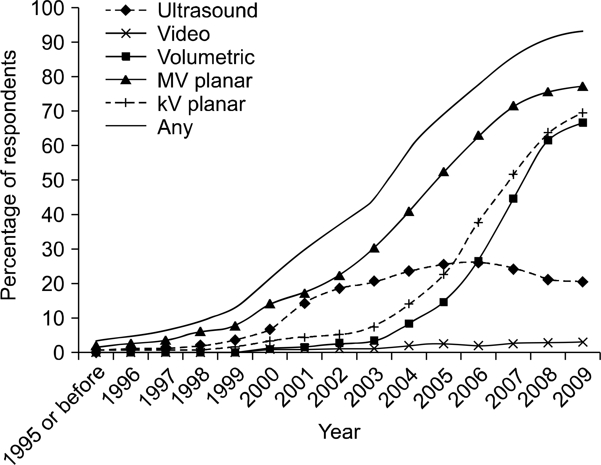
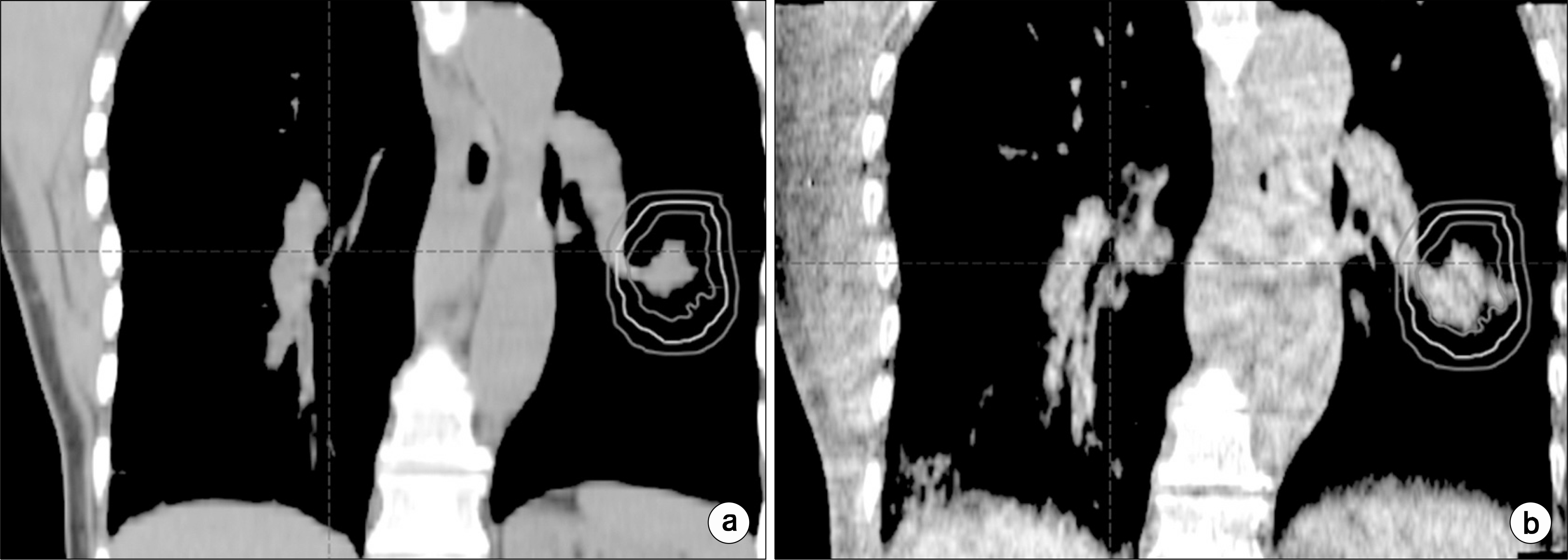
Fig. 3.
How the isocenter is determined for EPID system (Modified from page 3 of Varian Customer Technical Bulletine).23) Tolerance distance of Varian Exact Arm (E-Arm) is recommended to be 3 mm.
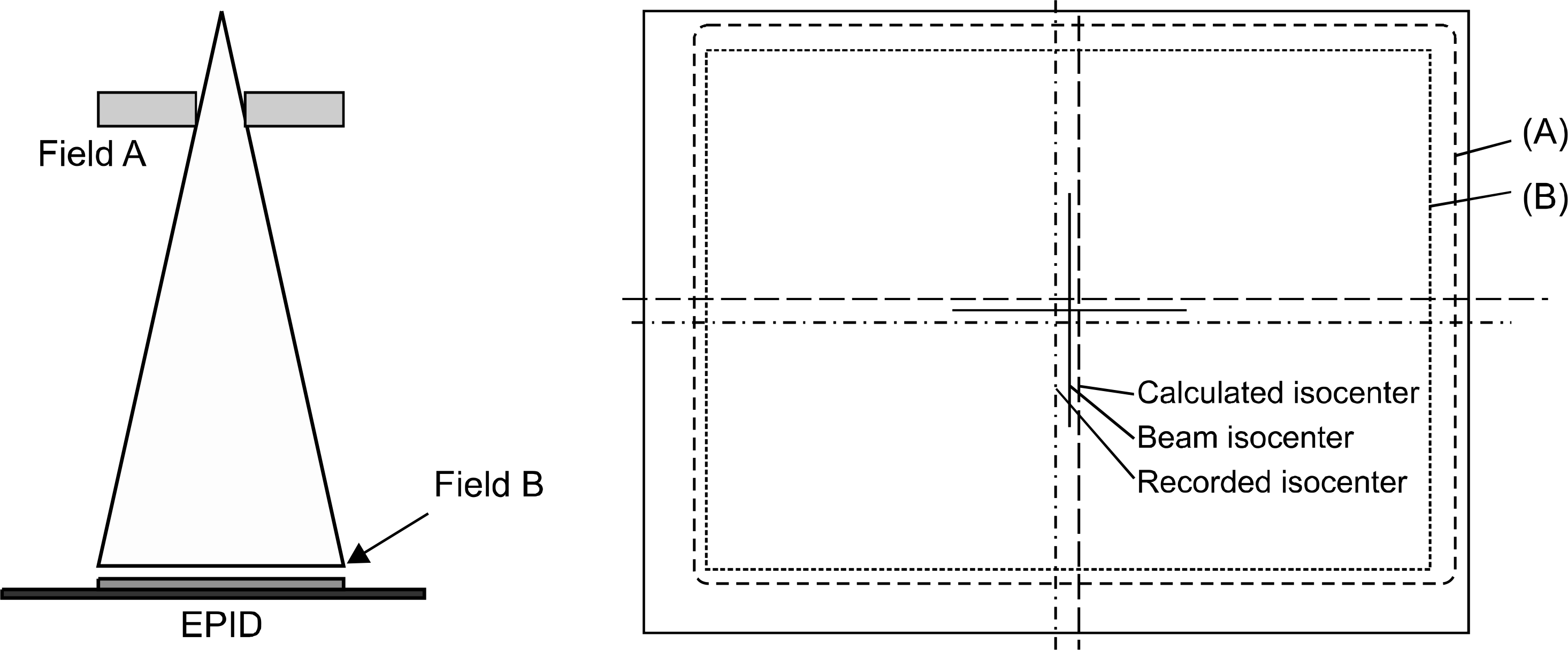
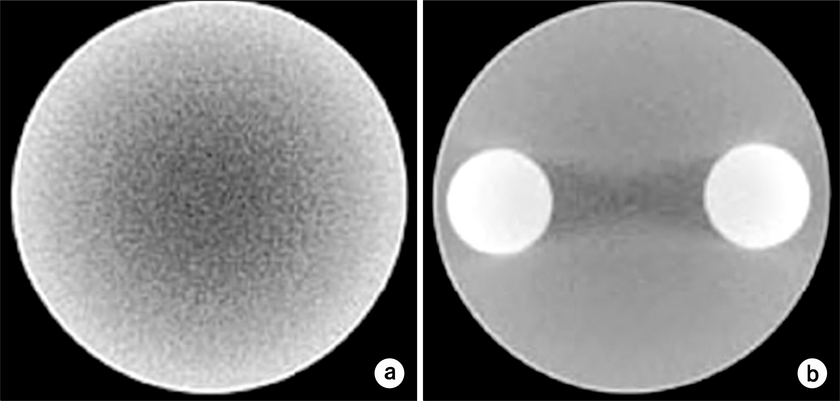
Fig. 4.
Effect of misalignment of the imaging center and the radiation center Misalignment of Digital Graticule can lead setup error. Setup accuracy relies on the machine tolerance.
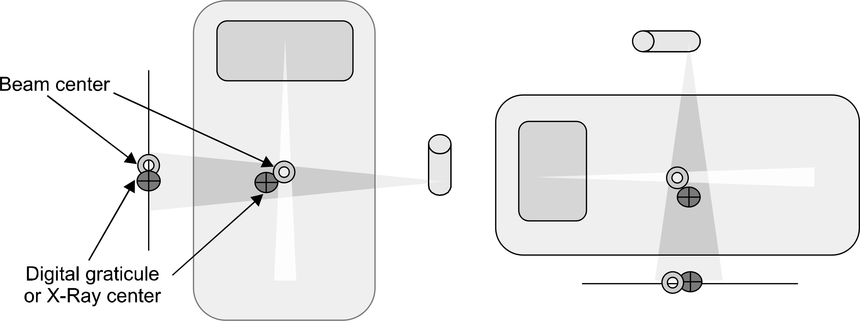
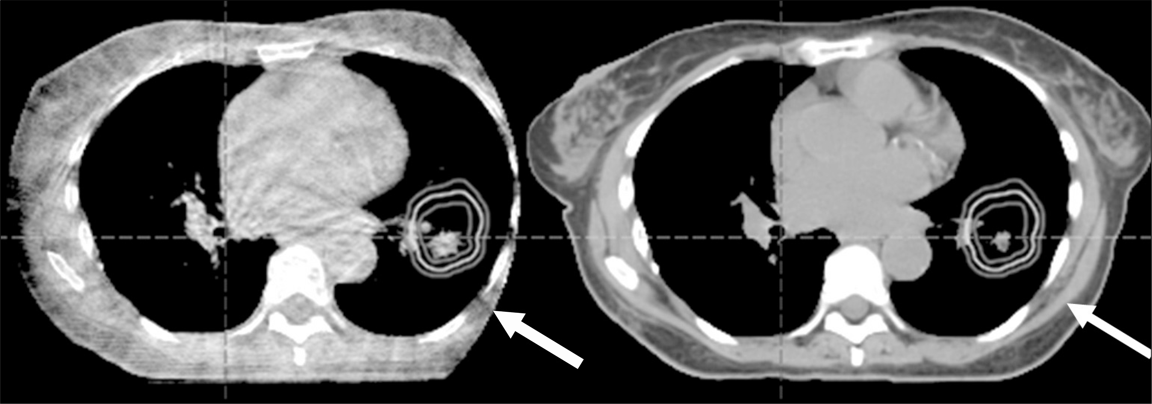
Fig. 6.
Artifacts of CBCT images from surrounding materials: (A and B) cupping and streaks due to hardening and scatter. Reprinted from Figure III-B-1 of reference 4. This figure is recited from Fig. 7, 8, p. 274, The Modern Technology of Radiation Oncology, Volume 2, J. Van Dyk (Ed.) Medical Physics Publishing.
 XML Download