Journal List > J Cardiovasc Imaging > v.26(2) > 1097081

Kim, Ann, Kim, Park, Won, and Lee: A Case of Unroofed Coronary Sinus with Coronary Sinus Orifice Atresia: Use of Multimodality Imaging
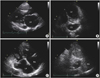
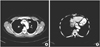
A 44-year-old woman visited our clinic because of sudden onset pain in both lower extremities. After admission, computed tomography (CT) was performed and revealed occlusion of both popliteal arteries and renal infarction in the left kidney. Transthoracic echocardiography (TTE) revealed rheumatic mitral stenosis (Figure 1A). In the apical view of TTE and on transesophageal echocardiography, we identified a huge dilated coronary sinus (CS) (Figure 1B) and detected large defect between the dilated CS and left atrium (LA) (Figure 1C, Movie 1). On contrast echocardiography with agitated saline, the right atrium (RA) was filled with air bubbles, but not the dilated CS, and then the right ventricle was filled with air bubbles later (Figure 1D). This finding showed no persistent left superior vena cava (PLSVC), which was supported by the CT imaging finding (Figure 2A). On CT, a huge dilated CS was observed, showing no communication with the RA, which suggested CS orifice atresia (Figure 2B). On real-time three-dimensional (3D) echocardiography in the en face view of the LA, an ovoid-shaped defect was found between the CS and the LA, posterior to the mitral valve (Figure 3A). After horizontal rotation of the image, no communication between the CS and the RA was visible, which suggested CS orifice atresia (Figure 3B). The patient was discharged after stabilization.
Unroofed coronary sinus (UCS) is a spectrum of cardiac anomalies in which part or all of the common wall between the CS and the LA is absent.1) Seventy-five percent of the UCS is associated with PLSVC.1) Atresia of the RA orifice of the CS is rare. Most cases of CS orifice atresia are associated with an alternative exit for coronary venous blood return, such as a small PLSVC, large thebasian vein, or CS canal defect, like in the present case.2) Myocardial ischemia is unlikely as long as an alternate exit exists for CS blood.3) Multimodality imaging such as, TTE, transesophageal echocardiography, contrast echocardiography, 3D echocardiography, CT, or magnetic resonance imaging is needed to diagnose this anomaly, like in the present case. In case of operation, the flow of CS blood should be retained. Deaths due to myocardial ischemia after ligation of a PLSVC were reported.4)
Figures and Tables

Figure 1

Transthoracic echocardiography revealed mitral stenosis and unroofed coronary sinus. (A) Doming of the anterior mitral leaflet and limited motion of the posterior mitral leaflet were demonstrated. (B) In the apical 4-chamber view with tilting, a huge dilated CS was identified. (C) With counterclockwise rotation of the probe, a 22-mm large defect between the dilated CS and the LA was detected. (D) Agitated saline was injected via the left antecubital vein. On contrast echocardiography, the RA was filled with air bubbles, but not the dilated CS, and then the right ventricle was filled with air bubbles later. This finding showed no persistent left superior vena cava. There was no negative jet in the RA, which raised the possibility of CS orifice atresia. CS: coronary sinus, LA: left atrium, LV: left ventricle, RA: right atrium, RV: right ventricle.

jcvi-26-103-g001
Figure 2

Computed tomography. (A) On computed tomography imaging, there was no persistent left superior vena cava (arrow). (B) A huge dilated CS was observed, showing no communication with the RA, which suggested CS orifice atresia. Ao: aorta, CS: coronary sinus, LV: left ventricle, RA: right atrium.

jcvi-26-103-g002
Figure 3

Three-dimensional echocardiography. (A) In the en face view of the LA, an ovoid-shaped defect (asterisk) was found between the CS and the LA, posterior to the mitral valve. (B) After horizontal rotation of the image, the bottom of the CS and the blinded pouch were visible through the defect. No communication between the CS and the right atrium was visible, which suggested CS orifice atresia. CS: coronary sinus, LA: left atrium.

jcvi-26-103-g003

Notes

Conflict of Interest The authors have no financial conflicts of interest.

References

1. Kong PK, Ahmad F. Unroofed coronary sinus and persistent left superior vena cava. Eur J Echocardiogr. 2007; 8:398–401.
crossref
2. Shum JS, Kim SM, Choe YH. Multidetector CT and MRI of ostial atresia of the coronary sinus, associated collateral venous pathways and cardiac anomalies. Clin Radiol. 2012; 67:e47–e52.
crossref
3. Santoscoy R, Walters HL 3rd, Ross RD, Lyons JM, Hakimi M. Coronary sinus ostial atresia with persistent left superior vena cava. Ann Thorac Surg. 1996; 61:879–882.
crossref
4. Yokota M, Kyoku I, Kitano M, et al. Atresia of the CS orifice. Fatal outcome after intraoperative division of the drainage left superior vena cava. J Thorac Cardiovasc Surg. 1989; 98:30–32.

SUPPLEMENTARY MATERIAL

Movie 1

On transesophageal echocardiography in 110° view, a defect was found between the dilated coronary sinus and the left atrium, and very slow flow through the defect was observed with spontaneous echo contrast.
TOOLS
Similar articles