Dear Editor:
It has been well known that FK 506 could decrease the atopic inflammation through controlling T-cells and their cytokines1. However, the effect of FK 506 on wounds has scarcely been investigated although FK 506 has been widely used in treating moderate to severe atopic dermatitis, where various minor wound occurs because of scratch owing to itchy. Therefore, we investigated the effect of FK 506 on wound healing with an in vitro study of outer root sheath keratinocytes (ORSKs) because ORSK is a key factor in cutaneous wound healing2.
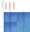
Human ORSKs were prepared using a method described previously with minor modifications3. We used the Ez-cytox® assay kit (ITSBIO, Seoul, Korea) to measure the cell proliferation. Cultured ORSKs were incubated for 4 days in 96-well microtiter plates in a final volume of 100 µl/well culture medium and various concentrations of FK 506. And then tetrazolium salt (10 µl) was added to each well and the absorbance was checked using a microtiter plate reader at 420∼480 nm. For migration assay, cell monolayer in culture dish divided by scratching with a p200 pipet tip. We added 104 nM FK 506 to the medium and created 10 different markings to be used as reference points. An initial photograph was taken, and additional images were obtained after 48 hours incubation. The width of the gap was measured using the TOMORO ver. 3.5 Scope Eye image analyzer program (Techsan Digital Imaging, Seoul, Korea). Cytokeratins 6, 16, and 17, as well as transforming growth factor-beta 1, 2 (TGF-β1, 2) have an important role in proliferation, and migration of keratinocytes. We assayed the cytokines using reverse transcription-polymerase chain reaction after treatment of FK 506. Data were evaluated using Student's t-test. ORSK proliferation in FK 506-treated groups (102 and 104 nM) increased to 103.7%±3.8% and 110%±9.0% of the control group (100%), respectively. It was not significant statistically in eight independent experiments (Fig. 1A). ORSK migration increased significantly in response to FK 506 (p<0.05). ORSKs treated with FK 506 occupied up to 75.8%±5.5% of the initial gap, whereas the control cells were about 9.4%±1.6% in three independent experiments (Fig. 1B).
The TGF-β1 expression increased 48 hours after treatment with 104 nM FK 506 (Fig. 2A). Cytokeratin 6, 16, and 17 mRNA levels did not change in any of the FK 506-treated groups (Fig. 2B).
10∼104 nM FK 506 did not inhibit proliferation of normal human epidermal keratinocytes (NHEK)45. In the present study, FK 506-treated ORSK proliferation was not significant statistically. Cytokeratin 6, 16, and 17 are highly expressed in the hyperproliferative epidermis in wound healing6. However their levels did not increase in FK 506-treated ORSKs, which was consistent with the finding that cell proliferation did not increase in our study. Thus, it appears that FK 506 had no effect on ORSK or NHEK proliferation.
ORSK migration increased significantly in response to 104 nM FK 506. Keratinocyte migration, rather than proliferation, plays a major role during re-epithelialization of epidermal defects7. In this study, TGF-β1 was detected 48 hours after treating the ORSKs with 104 nM FK 506. TGF-β1 plays a critical role in keratinocyte migration 78. Thus, the increase in ORSK migration in our study may be partially due to TGF-β1 expression. The cytoskeleton generally provides the strength for keratinocytes move and lamellopodial crawling during re-epithelialization. In particular, cytokeratin 6, 16, and 17 were induced in ORSKs at the wound's edge8. However, cytokeratin 6, 16, and 17 expression did not change in FK 506-treated ORSKs.
In conclusion, we showed that FK 506 did not inhibit ORSK proliferation but increased ORSK migration, which could have been partially due to TGF-β1 expression.
Figures and Tables
Fig. 1
(A) Proliferation assay for tacrolimus (FK 506)-treated human outer root sheath keratinocytes (ORSKs) using Ez-cytox®. A dose dependent increase was detected but it was not statistically significant. Results are shown as mean±standard deviation (SD) of eight independent experiments. (B) ORSKs treated with FK 506 occupied up to 75.8%±5.5% of the initial gap, whereas the control cells were about 9.4%±1.6% (p<0.05). Results are shown as mean±SD of three independent experiments.

Fig. 2
(A) Transforming growth factor-beta (TGF-β1) was highly expressed 48 hours after treatment of human outer root sheath keratinocytes (ORSKs) with 104 nM tacrolimus (FK 506), whereas no change was detected in the control, 102 or 103 nM FK 506-treated groups. (B) No change in cytokeratin 6, 16, and 17 mRNA expression after ORSKs were treated with FK 506. CYP: cyclophilin A, KRT: cytokeratin.

ACKNOWLEDGMENT
This study was funded by Institute of Medical Science Research of Dankook University Medical Center in 2012.
References
1. Park CW, Lee KY, Lee EH, Lee CH. The effects of cyclosporin A and FK-506 on the cytokine production of lymphocytes in atopic dermatitis. Ann Dermatol. 1996; 8:98–106.

2. Myers SR, Leigh IM, Navsaria H. Epidermal repair results from activation of follicular and epidermal progenitor keratinocytes mediated by a growth factor cascade. Wound Repair Regen. 2007; 15:693–701.

3. Kwack MH, Sung YK, Chung EJ, Im SU, Ahn JS, Kim MK, et al. Dihydrotestosterone-inducible dickkopf 1 from balding dermal papilla cells causes apoptosis in follicular keratinocytes. J Invest Dermatol. 2008; 128:262–269.

4. Kaplan A, Matsue H, Shibaki A, Kawashima T, Kobayashi H, Ohkawara A. The effects of cyclosporin A and FK506 on proliferation and IL-8 production of cultured human keratinocytes. J Dermatol Sci. 1995; 10:130–138.

5. Karashima T, Hachisuka H, Sasai Y. FK506 and cyclosporin A inhibit growth factor-stimulated human keratinocyte proliferation by blocking cells in the G0/G1 phases of the cell cycle. J Dermatol Sci. 1996; 12:246–254.

6. Sarret Y, Woodley DT, Grigsby K, Wynn K, O'Keefe EJ. Human keratinocyte locomotion: the effect of selected cytokines. J Invest Dermatol. 1992; 98:12–16.


 PDF
PDF ePub
ePub Citation
 Print
Print


 XML Download