Journal List > Korean Circ J > v.48(3) > 1093984

Kuwabara, Ishimura, Ishiwata, and Ohno: Isolated Cardiac Sarcoidosis Presenting with Stroke
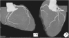
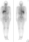
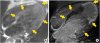
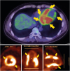
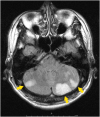
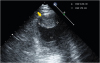
A 42-year-old man without past medical history presented to our hospital with temporary right sided hemiparalysis and dysarthria. Brain magnetic resonance imaging (MRI) showed multiple cerebrovascular infarctions (Figure 1). Electrocardiography showed sinus rhythm with frequent premature ventricular contractions (PVCs), but no atrial ventricular block. Echocardiography showed partial dyskinesis at the apical lesion and a significant left ventricular (LV) thrombus (Figure 2). Coronary computed tomography angiography (CTA) showed normal coronary arteries (Figure 3). The thrombus became small 4 weeks after anticoagulation therapy with warfarin, but it remained. Therefore, we could not conduct cardiac tissue biopsy because of the additional stroke risks. Gallium-67 scintigraphy showed no specific lesion (Figure 4). However, cardiac MRI showed multiple focal late gadolinium enhancement (LGE) in left ventricle (Figure 5B) where T2 star weighted image (T2WI) also showed pale high intensity (Figure 5A). It suggested active inflammation. Fasting 18F-fluorodeoxyglucose positron emission tomography (PET) after carbohydrate restriction showed multiple increased cardiac uptake lesions (Figure 6). However, apical lesions showed low uptake, which suggests scar lesions. CTA showed normal coronary arteries but cardiac MRI and PET showed multiple focal lesions, which suggests perfusion-metabolism mismatch. In these results, we diagnosed him with isolated cardiac sarcoidosis and started treatment with 30 mg/day (0.5 mg/kg/day) of prednisone.1) The cardiac function improved by echocardiography, the number of PVCs became half in 24-hour Holter electrocardiogram, and follow-up PET image showed less cardiac uptake 3 months after treatment.
Some cases of systematic sarcoidosis presenting with stroke or heart failure were reported,2)3) but our case is an isolated cardiac sarcoidosis presented with stroke. Cardiac MRI and PET are useful tools to diagnose isolated cardiac sarcoidosis.4) We should consider isolated cardiac sarcoidosis as a cause of stroke. A study showed that patients with definite cardiac sarcoidosis treated with steroids had better outcome than those with probable cardiac sarcoidosis without treatment.5) This case report suggests that early treatment for isolated cardiac sarcoidosis could be effective.
Figures and Tables

Figure 1
Brain magnetic resonance image. This image shows multiple cerebrovascular infarctions especially in the cerebellum.
kcj-48-236-g001
Figure 2
Echocardiography shows LV thrombus. The thorombus is 2.64×1.74 cm in the LV apical lesion.
LV = left ventricular.
kcj-48-236-g002
Figure 3
Coronary CTA. This image shows normal coronary arteries.
CTA = computed tomography angiography.
kcj-48-236-g003
Figure 4
Gallium-67 scintigraphy. This image shows no specific lesion in the body.
kcj-48-236-g004
Figure 5
Cardiac MRI image. (A) T2WI and (B) LGE image. There are multiple focal LGEs in left ventricle (B) where T2WI also shows pale high intensity with unclear margin. It suggests active inflammation.
LGE = late gadolinium enhancement; MRI = magnetic resonance imaging; T2WI = T2 star weighted image.
kcj-48-236-g005
Figure 6
Fasting 18F-fluorodeoxyglucose PET. The image shows multiple increased cardiac uptake lesions, especially at the anterior, basal lateral, and inferior LV lesions. The contrast is clear, and the lesions are very similar to LGE lesions. However, apical lesions show low uptake, which suggests scar lesions.
LGE = late gadolinium enhancement; LV = left ventricular; PET = positron emission tomography.
kcj-48-236-g006

Notes

Conflict of Interest The authors have no financial conflicts of interest.

Author Contributions

  • Conceptualization: Kuwabara M, Niitsu Ishimura R, Ishiwata S, Ohno M.

  • Data curation: Kuwabara M, Niitsu Ishimura R.

  • Formal analysis: Kuwabara M, Niitsu Ishimura R.

  • Investigation: Kuwabara M.

  • Methodology: Kuwabara M, Ishiwata S, Ohno M.

  • Project administration: Kuwabara M.

  • Resources: Kuwabara M, Niitsu Ishimura R.

  • Software: Kuwabara M, Niitsu Ishimura R.

  • Supervision: Kuwabara M, Niitsu Ishimura R.

  • Validation: Kuwabara M, Niitsu Ishimura R, Ishiwata S, Ohno M.

  • Visualization: Kuwabara M.

  • Writing - original draft: Kuwabara M.

  • Writing - review & editing: Kuwabara M, Niitsu Ishimura R, Ishiwata S, Ohno M.

References

1. Sadek MM, Yung D, Birnie DH, Beanlands RS, Nery PB. Corticosteroid therapy for cardiac sarcoidosis: a systematic review. Can J Cardiol. 2013; 29:1034–1041.
2. Brown MM, Thompson AJ, Wedzicha JA, Swash M. Sarcoidosis presenting with stroke. Stroke. 1989; 20:400–405.
crossref
3. White J, Sutton T, Kerr A. Isolated primary cardiac sarcoidosis: MRI diagnosis and monitoring of treatment response with cardiac enzymes. Circ Heart Fail. 2010; 3:e28–9.
4. Isobe M, Tezuka D. Isolated cardiac sarcoidosis: clinical characteristics, diagnosis and treatment. Int J Cardiol. 2015; 182:132–140.
crossref
5. Takaya Y, Kusano KF, Nakamura K, Ito H. Comparison of outcomes in patients with probable versus definite cardiac sarcoidosis. Am J Cardiol. 2015; 115:1293–1297.
crossref
TOOLS
ORCID iDs

Masanari Kuwabara
https://orcid.org/0000-0002-6601-4347

Similar articles