

 PDF
PDF ePub
ePub Citation
 Print
Print


INTRODUCTION
Atrial fibrillation (AF) is the most common cardiac arrhythmia. The prevalence of AF increases with age, and affects 3% to 5% of people over 65. AF increases the risk of ischemic stroke by five times and causes approximately 15% to 20% of ischemic strokes.1) AF is also associated with an increased risk of heart failure and mortality. Although several risk factors of AF have been identified, including high blood pressure, heart failure, diabetes, ageing, hyperthyroidism, and heart disease, the pathogenesis of AF remains poorly understood.
Recent studies revealed that serum parathyroid hormone (PTH) levels are higher in subjects with AF than in controls.2)3)4) Another prospective study found that a higher serum level of phosphorus was associated with an increased risk of AF.5) Also, PTH excess was associated with major risk factors for the development and perpetuation of AF, including hypertension, myocardial dysfunction, and heart failure.6)7)8) These findings suggest that serum PTH levels might be related with AF.
However, these previous studies had limitations due to the small number of patients enrolled and the independent effect of PTH on AF prevalence was not analyzed. To our knowledge, there has not been a population based study on the association between serum PTH and AF. Therefore, the aim of this study was to investigate the association between serum PTH levels and the prevalence of AF in a population based study.
METHODS
Study population
The Dong-gu study is an ongoing prospective population-based study that was designed to investigate risk factors for chronic disease among 9,260 subjects aged 50 years and older in an urban elderly population.9) Baseline examination of the Dong-gu study was conducted between April 2007 and July 2010 in the Dong-gu district of Gwangju Metropolitan City in Korea. After the exclusion of 228 participants who had incomplete data, a total of 9,007 subjects (3,606 men and 5,401 women) were included in the present study. All participants provided informed consent, and the study was conducted in accordance with the guidelines in the Declaration of Helsinki. The study was approved by the Institutional Review Board of Chonnam National University Hospital (approval number: I-2008-05-056).
Covariates
Body weight and body composition were measured while the subjects were wearing indoor clothing or a light gown without shoes using a calibrated Inbody 520 (Biospace Co., Seoul, Korea). Height was measured to the nearest 0.1 cm. Subjects were categorized based on smoking status as current smokers or nonsmokers (including ex-smokers), and based on alcohol intake as nondrinkers or current drinkers. Regular exercise was categorized as irregular or regular based on the weekly frequency of recreational activity and exercise. After the subjects had rested for at least 5 minutes in a sitting position, blood pressure was measured on the right upper arm using a mercury sphygmomanometer (Baumanometer; W.A. Baum Co., Inc., Copiague, NY, USA) with an appropriately sized cuff. The first appearance (phase I) and disappearance (phase V) of the Korotkoff sounds were used to determine systolic blood pressure (SBP)and diastolic blood pressure (DBP), respectively, which was recorded to the nearest 2 mmHg. SBP and DBP were measured three times at one-minute intervals, and the average values were used in our analysis. All participants underwent at least 10 hours of overnight fasting before blood samples were obtained from an antecubital vein. Blood sampling was performed at a pre-fixed morning time between 8 and 9 AM. Serum was separated on-site and stored at −70°C until analyzed. Serum total cholesterol, high-density lipoprotein (HDL) cholesterol, triglycerides, and fasting blood glucose levels were measured using enzymatic methods. All samples were analyzed using an automatic analyzer (Model 7600 Chemical Analyzer; Hitachi Ltd., Tokyo, Japan). C-reactive protein (CRP) levels were determined by particle-enhanced immunonephelometry using a BN II nephelometer (Dade Behring, Marburg, Germany). Kidney function was assessed by the estimated glomerular filtration rate (eGFR), which was calculated using the Modification of Diet in Renal Disease (MDRD) formula. Chronic kidney disease was defined as an eGFR <60 mL/min.
Serum PTH measurement
Concentrations of PTH were measured by a chemiluminescent microparticle immunoassay (ARCHITECT i2000; Abbott, Abbott Park, IL, USA). The coefficient of variation for the total analytic precision of this assay was ≤9%. The lower detection limit of this assay was 1.0 pg/mL for PTH.
Assessment of AF
All participants underwent a 12-lead electrocardiogram (ECG). The ECGs were recorded using the same HP-PageWriter 200 M1771A (Hewlett Packard, Andover, MA, USA). AF was diagnosed if AF or atrial flutter was present on an ECG obtained from a baseline survey. The ECGs were initially analyzed by a computer-based ECG interpretation program (Philips 12-lead algorithm). All potential cases of AF were determined by two cardiologists.
Statistical analyses
The data are presented as the mean value and standard deviation for normally distributed continuous variables and proportions for categorical variables. Differences across quartiles of PTH were assessed by linear trends in the χ2 test and analysis of variance test, as appropriate. Multiple logistic regression analysis was used to evaluate the relationship between the quartiles of PTH and AF, adjusting for potential confounders. We included all variables known to be confounder variables in previous studies as independent variables in the multivariate analysis. However, the levels of calcium and phosphorus that could mediate the association between PTH levels and AF were not included in the independent variables. The following cardiovascular risk factors were included as covariates: age, gender, body mass index (BMI), smoking, alcohol intake, physical activity, medication for hypertension, medication for diabetes, medication for dyslipidemia, SBP, DBP, fasting glucose, total cholesterol, HDL cholesterol, log-transformed triglyceride, aspartate aminotransferase (AST), alanine aminotransferase (ALT), and log-transformed values of high-sensitivity CRP and eGFR. The adjusted prevalence ratio was the predicted probability calculated from the logistic regression model using the “margins” command with the “atmeans” option in Stata. All statistical analyses were performed using Stata version 14 (StataCorp, College Station, TX, USA).
RESULTS
The general characteristics for the Dong-gu study participants by quartile of serum PTH level are shown in Table 1. The mean age of the study participants was 65.2 (±8.6) years and 40.0% were males. The median serum PTH level was 39.5 pg/mL (interquartile range (IQR), 30.6–50.8), which was significantly higher in patients with AF than without AF (49.1 [IQR, 37.4-71.5] vs. 39.3 [IQR, 30.5-50.6] pg/mL; p<0.001) (Figure 1). Subjects in the highest quartile of serum PTH were more likely to be older, female, non-smokers, non-drinkers, and physically inactive. In addition, they had significantly higher mean values for BMI, SBP, and DBP, but lower values for fasting glucose, total cholesterol, triglyceride, and eGFR.
Table 1
General characteristics of the study population (n=9,007) according to serum PTH level
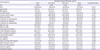
Data shown are number (%) or mean±standard deviation.
ALT = alanine aminotransferase; AST = aspartate aminotransferase; BMI = body mass index; CRP = C-reactive protein; DBP = diastolic blood pressure; eGFR = estimated glomerular filtration rate; HDL = high-density lipoprotein; PTH = parathyroid hormone; SBP = systolic blood pressure.
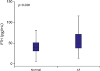
Figure 1
Serum levels of PTH in patients with normal sinus rhythm and AF. The median and IQR of serum PTH was 39.5 pg/mL (IQR, 30.6–50.8), which was significantly higher in patients with AF than without AF (p<0.001).
AF = atrial fibrillation; IQR = interquartile range; PTH = parathyroid hormone.
The prevalence of AF was 2.41% in males and 0.9% in females and increased with advanced age in both genders. The prevalence of AF increased with increasing PTH quartile in both males (1.6%, 1.9%, 2.3%, and 4.2% in the lowest, second, third, and highest quartiles, respectively; p<0.001) and females (0.2%, 0.2%, 1.1%, and 1.7%; p<0.001). There was no sex difference in the association between PTH level and the prevalence of AF (p for interaction=0.10). The prevalence of AF increased with increasing PTH quartile even after adjusting for age and gender (0.8%, 0.9%, 1.6%, and 2.8%; p<0.001) (Figure 2). The association between serum PTH and prevalence of AF is shown in Table 2. Serum PTH levels were positively associated with the risk for AF after adjustment for age and sex (odds ratio [OR] for the second quartile, 1.09; 95% confidence interval [CI], 0.57–2.08; OR for the third quartile, 2.03; 95% CI, 1.14–3.60; and OR for the fourth quartile, 3.65; 95% CI, 2.14–6.22). After additional adjustment for cardiovascular risk factors, this association was slightly attenuated and remained significant (OR for the second quartile, 1.04; 95% CI, 0.54–1.99; OR for the third quartile, 1.95; 95% CI, 1.09–3.48; OR for the fourth quartile, 3.34; 95% CI, 1.93–5.78). Multivariate analysis revealed that increasing age, female gender, high SBP, high DBP, and increasing serum PTH level were associated with higher prevalence of AF (Table 3).
Figure 2
The prevalence of AF according to PTH quartiles adjusted by age and gender. Adjusted prevalence was calculated by logistic regression adjusted for age and sex. The prevalence of AF even after age and gender adjustment increased with increasing PTH quartile (0.8%, 0.9%, 1.6%, and 2.8% in the lowest, second, third, and highest quartiles, respectively; p<0.001).
AF = atrial fibrillation; PTH = parathyroid hormone.
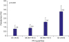
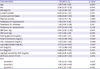
Table 2
ORs for AF by PTH quartiles in 9,007 participants from the Dong-gu study

AF = atrial fibrillation; ALT = alanine aminotransferase; AST = aspartate aminotransferase; BMI = body mass index; CI = confidence interval; CRP = C-reactive protein; DBP = diastolic blood pressure; eGFR = estimated glomerular filtration rate; HDL = high-density lipoprotein; OR = odds ratio; PTH = parathyroid hormone; SBP = systolic blood pressure.
*Adjusted for age, sex, BMI, smoking, alcohol intake, physical activity, medication for hypertension, medication for diabetes, mediation for dyslipidemia, SBP, DBP, fasting glucose, total cholesterol, HDL cholesterol, AST, ALT, log-transformed triglyceride, log-transformed values of high-sensitivity CRP, and eGFR.
Table 3
Multivariate analysis for association with the prevalence of AF
AF = atrial fibrillation; ALT = alanine aminotransferase; AST = aspartate aminotransferase; BMI = body mass index; CI = confidence interval; CRP = C-reactive protein; DBP = diastolic blood pressure; eGFR = estimated glomerular filtration rate; HDL = high-density lipoprotein; OR = odds ratio; PTH = parathyroid hormone; SBP = systolic blood pressure.
DISCUSSION
AF is the most common form of sustained arrhythmia in clinical practice and occurs in approximately 1.5% of adults. AF is particularly important because it is associated with increased morbidity and mortality.1) Also, AF is a major risk factor for cerebral embolism. Thus, identifying contributing factors for the development of AF is critical. Numerous risk factors have been suggested to be associated with an increased prevalence of AF, including high blood pressure, heart failure, diabetes, ageing, hyperthyroidism, and heart disease. Nonetheless, the cause of AF in a substantial number of patients remains unexplained.
The median serum level of PTH has been found to be higher in patients with AF than in patients with a normal sinus rhythm. PTH plays a key role in calcium homeostasis and its release is triggered by a decrease in serum calcium levels, acting on bone, kidney, and intestine. Beyond these classical targets, PTH also acts as a cardiac hormone, a vasodilatory substance, and a regulator of smooth muscle proliferation.10)11) An increased serum level of PTH was associated with hypertension, disturbances in the renin-angiotensin-aldosterone system, and cardiac arrhythmia, as well as structural and functional alterations in the vascular wall.12)13) Previous population-based studies have reported that higher PTH levels were associated with higher levels of SBP and DBP.14) Also, PTH infusion in healthy individuals raised blood pressure.15) In the present study, treatment history of hypertension was significantly increased according to the increment of PTH quartiles. History of hypertension was marginally associated with the risk of AF even after covariate adjustment. Hypertension is a well-known risk factor for the development of AF and its complications. Hypertension leads to atrial and ventricular pressure overload and arterial and atrial stiffness, resulting in increased atrial workload and distension. Therefore, hypertension contributes to the elevated serum levels of PTH and the development of AF.
Another potential mechanism for the association between AF and PTH is that AF itself might increase PTH levels. Messenger RNA expression of PTH-related proteins in the heart is increased by physiologic maneuvers that increase atrial or ventricular workload and distention.10) Loss of atrial contraction during AF causes atrial volume and pressure overload leading to atrial stretch. Also, the pathomechanism of AF involves atrial ischemia, infiltration, and inflammation, which promote local release of PTH.16) This mechanism suggests that PTH release into the circulation could increase during AF. Increased levels of PTH may perpetuate AF by direct effects on cardiomyocytes. PTH can induce myocardial hypertrophy and increase heart rate and automaticity, and thus increase the risk of AF.11)17) McCarty et al.18) demonstrated that PTH-induced activation of phospholipase C may increase arrhythmia by the generation of inositol-1,4,5-triphosphate, which has been associated with increased arrhythmogenicity.
Consistent with the present study, previous studies revealed that patients with AF presented higher serum levels of PTH than patients with a normal sinus rhythm. The increased serum level of calcium with increasing PTH quartile in the present study supports the explanation that an increased serum level of PTH is probably a response to AF. However, further studies are needed to evaluate the causal relationship between PTH and AF.
PTH excess increases intracellular calcium in target tissues and is associated with hypertension, arterial stiffness, left ventricular hypertrophy, and heart failure.12) Recent clinical studies revealed that PTH excess was significantly associated with an increased risk for the development of cardiovascular disease and mortality.19)20) Also, PTH excess was positively linked with the risk and severity of heart failure.4)6) Patients with PTH excess were more likely to be older and physically inactive and had a higher prevalence of comorbidities, resulting in an increased risk of cardiovascular disease. Population based studies have shown that PTH excess was associated with major risk factors for the development of AF, including hypertension, left ventricular hypertrophy, myocardial dysfunction, left ventricular mass, and heart failure.7)8)12) In the present study, subjects with higher serum PTH were more likely to be older, female, non-smokers, non-drinkers, and physically inactive. Also, they had a higher mean BMI and higher prevalence of hypertension. These demographic features might contribute to the higher prevalence of AF in patients with higher PTH levels. The Dong-gu study is an ongoing prospective population-based study and all participants are being examined for the development of cardiovascular disease using echocardiograms, ECGs, and laboratory examinations for 5 years after the initial enrollment. The Dong-gu study is expected to show the temporal relationship between serum levels of PTH and cardiovascular disease including heart failure as well as AF.
Some limitations of the present study need to be addressed. First, the serum level of PTH was not analyzed according to the AF type as either paroxysmal or persistent. Previous studies demonstrated that patients with persistent AF had significantly higher PTH levels than patients with paroxysmal AF.2)3) Follow-up data from the Dong-gu study are expected to discriminate the association of PTH according to AF type. Second, the serum level of vitamin D was not incorporated into the multivariate logistic model. Vitamin D is another key factor involved in controlling calcium and phosphate homeostasis. Although there is conflicting data about the relationship between serum levels of vitamin D and cardiovascular disease, higher vitamin D levels have been associated with a higher risk of cardiovascular mortality and heart failure.6)8)19)20) Third, the present study has a cross-sectional study design, which precludes determination of the causal relationship between PTH levels and AF prevalence, so further prospective studies are needed to reveal the cause and effect of this relationship. Fourth, only a single measurement of PTH level was performed and biologic variation in PTH is expected over time. Multiple measurements of the PTH level might reduce the incidental relationship between PTH levels and AF prevalence.
Higher levels of serum PTH were associated with higher prevalence of AF. Further studies are needed to determine whether the association is present in other populations and in a prospective study setting. Patients with higher PTH levels were older, physically inactive, obese, and had more comorbidities including hypertension. These features increased the risk of future cardiovascular disease. The Dong-gu study is expected to add evidence for the relationship between PTH levels and cardiovascular disease as well as AF.
 XML Download