

 PDF
PDF Citation
 Print
Print


INTRODUCTION
In recent years, an improved understanding of immune-modulatory signaling pathways in immune cells and the tumor microenvironment (TME) has led to rejuvenated interest in cancer immunotherapy. In particular, immune therapy targeting the immune checkpoint receptors such as cytotoxic T-lymphocyte-associated antigen 4 (CTLA-4), programmed cell-death 1 (PD-1), and programmed cell-death ligand 1 (PD-L1) are among the most promising approaches, having demonstrated clinical activity in a wide variety of tumors. This review will focus on the current evidence of immune checkpoint inhibitors in gynecologic malignancies, with specific emphasis on two of the most actively studied immune checkpoint receptors, CTLA-4 and PD-1 pathways.
Under physiologic conditions, immune checkpoints comprise several key inhibitory signals crucial for maintenance of self-tolerance. These inhibitory signals can be modulated by tumor cells to prevent the immune system from mounting an effective anti-tumor immune response [1]. The response of the immune system is initiated through antigen recognition by the T cell receptor (TCR) following antigen presentation by professional antigen-presenting cells (APCs) such as dendritic cells (DCs). It is regulated by a balance between co-stimulatory and inhibitory signals [234], leading to interactions between the TCR and antigenic tumor peptide bound to major histocompatibility complex (MHC) class I or II, resulting in T-cell mediated tumor destruction [5].
CTLA-4 is expressed exclusively on T cells and primarily counteracts the activity of the T cell co-stimulatory receptor, CD28 [6]. CTLA has a higher affinity to the B7 ligand on APCs than CD28, resulting in inhibition of T cell activation [1]. CTLA-4 functions as a signal dampener [78] with high-affinity ligands inducing higher levels of CTLA-4 [1]. The inhibitory role of the PD-1 pathway serves to enhance immune resistance in the TME by down-regulating the activity of T cells in peripheral tissues when bound to PD-L1, thus limiting collateral damage [1591011]. PD-L1 induction is via interferon-γ (IFN-γ), which is predominantly produced by T helper 1 (TH1) cells [1213].
Hence, by blocking the interactions mediating down regulation of T cell activity against tumors, immune checkpoint inhibitors aim to inhibit immune escape and improve recognition of tumors by the immune system. Here we describe the rationale for its use in several gynecologic tumors and review the literature for this promising approach.
IMMUNE CHECKPOINT INHIBITION IN ENDOMETRIAL CANCER
Endometrial cancer is the sixth most common malignancy in women worldwide, with majority of cases occurring in developed countries [14]. Historically, endometrial cancer has been divided into type I and type II based on clinicopathological characteristics. Type I tumors are of endometriod histology, generally associated with obesity, hormone receptor positivity, estrogen excess and a favorable prognosis. Type II tumors comprise primarily of serous and other histological subtypes, and have a worse clinical outcome [1516]. When diagnosed early, surgery and adjuvant therapy portend a favorable prognosis while those who are diagnosed at an advanced stage or with recurrent disease have few effective chemotherapeutic options [17].
1. Rationale for immune checkpoint inhibitors in endometrial cancer
Endometrial cancers can be molecularly classified into four distinct categories — an ultra-mutated group, hyper-mutated group, copy number low group and copy number high group [18]. The ultra-mutated group is characterized by extremely high mutation rates that harbors mutations in the exonuclease domain of polymerase ɛ (POLE) while tumors in the hyper-mutated group, consist of mainly mismatch repair (MMR) deficient tumors [18]. The copy number low group has lower mutation frequencies and consist primarily of the microsatellite stable (MSS) tumors. Similar to its ovarian counterpart, the serous-like tumors of the copy number high group have extensive somatic copy number aberrations (SCNA) with a low mutation rate [18].
The MMR pathway is a single strand break repair mechanism for DNA replication errors and inhibits recombination between non-identical (homologous) sequences [19]. It is critical for genomic stability and failure results in the microsatellite instability (MSI) and hyper-mutator phenotype [19]. Deficiency in MMR increases mutation rates up to 1,000-fold and is causally associated with the development of endometrial cancer (approximately 24% of cases), with MLH1 methylation being reported in 89% cases demonstrating MLH1/PMS2 immunohistochemistry (IHC) loss [2021]. Most MMR defects are caused by somatic epigenetic silencing of MLH1 but mutations in MLH1, MSH2, MSH6, MLH3, PMS1 or PMS2 can occur either somatically or inherited as germline mutations in Lynch syndrome [202122]. Lynch syndrome (also known as hereditary non-polyposis colon cancer [HNPCC]) is associated with early onset proximally sited colonic tumors and increased incidence of endometrial, stomach, small intestine, liver, brain, and urinary system cancers [21], commonly involves MLH1 (50%), MSH2 (39%) or MSH6 (7%) mutations with MLH3, PMS1 or PMS2 genes occasionally involved [22] and has a 40%–60% lifetime risk of developing endometrial and colorectal cancer [23]. Germline MSH6 mutations are associated with a high risk of endometrial cancer (71%) [24] and germline MSH2 mutations are at higher risk of developing extra-colonic cancers [25].
Aside from MSI, the loss of DNA catalytic and proofreading function in DNA POLE, is another important player in endometrial cancer tumorigenesis. POLE exonuclease domain mutations (EDMs) are found in 5%–8% of endometrial cancers [26], and have been shown to increase spontaneous mutations [27]. Several reports have suggested that ultra-mutated and hyper-mutated tumors may harbor more tumor-specific neoantigens resulting in increased amounts of tumor infiltrating lymphocytes (TILs) [2829], potentially making the ultra-mutated and hyper-mutated groups excellent candidates for immunotherapy.
High mutational load MSI and POLE endometrial cancers were observed to be associated with significantly increased predicted neoepitopes and CD3+/CD8+ TILs demonstrating that the neoantigen load is proportional to the mutational load [30]. POLE mutated tumors demonstrated neoantigen load 15 times higher compared to MSI tumors, which in turn demonstrated 7 fold higher neoantigen load compared with MSS tumors [30]. Furthermore, MSI and POLE endometrial cancers tended to overexpress PD-1/PD-L1 in both tumor cells as well as in the TME compared to their MSS counterparts [30].
This hypothesis was clinically validated in a pivotal phase II study using pembrolizumab, an anti-PD-1 inhibitor, in patients with previously treated metastatic carcinoma. Mismatch repair status was retrospectively assessed using a standard polymerase chain reaction (PCR)-based method [31]. Notably, objective immune related response rate (RR) and immune related progression free survival (PFS) rate were 40% and 78%, respectively, for MMR-deficient colorectal cancers and 0% and 11%, respectively, for MMR-proficient colorectal cancers [31]. Not surprisingly, responses in patients with MMR-deficient non-colorectal cancer (this cohort included 2 patients with endometrial cancer) reflected those of patients with MMR-deficient colorectal cancer — immune related RR 71% (5/7 patients); immune related PFS rate, 67% (4/6 patients) [31]. Whole-exome sequencing revealed a mean of 1,782 somatic mutations per tumor in MMR-deficient tumors, compared with 73 in MMR proficient tumors (p=0.007), and high somatic mutation loads were associated with prolonged PFS (p=0.02) following PD-1 blockade [31]. Intriguingly, membranous PD-L1 expression was noted in all MMR-deficient tumors and correlated with greater density of CD8-positive lymphoid cells [31].
Hence, the key question is how best to identify patients with endometrial cancers that are likely to harbor MMR-deficiency. When comparing the pathologic features of sporadic MSI-high endometrial carcinoma, sporadic endometrial cancer in women less than 50 years who were Lynch syndrome-negative, and Lynch syndrome-associated MSI-high, endometrial carcinoma, the sporadic group consisting of women less than 50 years with endometrial carcinoma and the sporadic MLH1 methylation group were almost entirely composed of tumors with endometrioid histology (41/42, 97.6% and 25/26, 96.2%, respectively) [32]. However, the histology of Lynch syndrome-associated cancers was more heterogeneous (86% endometrioid; 14% papillary serous, clear cell or malignant mixed Müllerian subtypes). Interestingly, all of the non-endometrioid tumors in this study occurred in patients with MSH2 mutations and the mean age of diagnosis in this group of patients was 46.4 years [32]. It is unclear if MSI-high endometrial carcinoma associated Lynch syndrome responds similarly to PD-L1 blockade as their colorectal counterparts (only 3/11 MSI-high Lynch syndrome responded to pembrolizumab compared to 6/6 non-Lynch syndrome MSI high). As we continue to evaluate the role of MSI in endometrial cancer, it is worthwhile to consider all patients diagnosed with endometrial cancer for MSI testing and germline testing for Lynch syndrome especially if diagnosed at a young age or have a family history of Lynch syndrome related cancers.
2. Clinical studies of PD-1/PD-L1 blockade in advanced endometrial cancer
An ongoing phase II study of advanced, pre-treated endometrial cancer harboring deficiency in MMR proteins has reported preliminary observations of overall response rate (ORR) of 55.6% (5/9) and a clinical benefit rate of 88.9% when treated with pembrolizumab. Of interest, one patient who achieved a sustained complete response (CR) for 17 months, had previously progressed through 3 prior lines of chemotherapy [33].
Preliminary results from the phase Ib KEYNOTE-028 (National Clinical Trial [NCT]02054806) cohort of 24 patients with MSI high advanced endometrial cancer, also suggest activity for pembrolizumab with tolerable toxicity. Recruited patients had PD-L1 expression in ≥1% of tumor or stromal cells by immunohistochemical staining. At a median of 69.9 weeks of follow-up, confirmed ORR was 13% (3/24), and another 13% (3/24) achieved stable disease (SD) [34]. We await the phase II KEYNOTE-158 trial (NCT02628067) that will better assess the efficacy of pembrolizumab in this setting.
IMMUNE CHECKPOINT INHIBITION FOR EPITHELIAL OVARIAN CANCER (EOC)
EOC is the fifth most common cause of cancer death in western women [35]. With no validated screening schedule, and paucity of symptoms at disease onset, more than 50% of ovarian cancers are diagnosed at an advanced stage [35]. Cytoreductive surgery integrated with platinum based chemotherapy has become the initial standard treatment [36373839]. However, approximately 70% of patients experience disease relapse after a varying disease-free interval. Pegylated liposomal doxorubicin [40], topotecan [41], and gemcitabine [42] are among the cytotoxic agents used in the platinum-resistant setting, with generally low RRs [404142]. Targeted approaches with anti-vascular endothelial growth factor (VEGF) antibody, bevacizumab [43], and olaparib, an inhibitor of the enzyme poly-(ADP ribose) polymerase inhibitor (PARPi), have also demonstrated improvement in outcomes for patients with advanced ovarian cancer [44]. More recently, there is an increasing body of evidence pointing to the intrinsic immunogenicity of advanced ovarian cancer with the presence of CD3+ TILs and a high CD8+/regulatory T cell ratio correlating with improved survival in ovarian cancer patients [4546], thus supporting the feasibility of an immunotherapeutic approach.
1. Rationale for immune checkpoint inhibitors in advanced EOC
In a previous study examining the prognostic relevance of the host immune response in ovarian cancer, intratumoral CD3+ T-cell infiltration was found to be an independent prognostic factor for improved outcomes in 186 patients with advanced ovarian cancers [45]. Conversely, infiltration of CD4+ CD25+ regulatory T-cells carried a worse prognosis [47]. More recently, intratumoral CD8+ TIL was observed to be superior compared to CD3 as a marker for immune cell infiltrates and correlates with improved clinical outcomes in a large cohort of ovarian cancer patients, particularly in the serous histology [48]. The presence of intratumoural TILs were also observed to be associated with BRCA mutation or epigenetic loss, suggesting a possible link to chromosomal instability [48]. In the serous histological subset of ovarian cancer, tumor infiltrating B cells (CD20+) appear to play an equally prominent role when they co-localized with T cells and display characteristics of APC [49]. The presence of both CD20+ B cells and CD8+ T cells in tumors has been associated with better prognosis than T cells alone [49]. However, the role of B cells in anticancer immunity remains controversial. Nevertheless, a recent study assessed co-localization patterns in the high grade serous histological subset and observed tumors containing CD8+, CD4+, and CD20+ TIL together with PCs were associated with markedly increased survival, with approximately 65% of patients alive at 10 years [50]. In addition, patients with plasma cell gene signatures were strongly associated with cytotoxic immune responses and improved survival [50]. Therefore, immunotherapeutic strategies that activate both lymphocyte subsets may have more potent and sustained antitumor effects.
Further evidence that an immunogenic TME may be associated with improved outcomes was observed in gene expression analysis of advanced EOC, that categorized the disease into four main molecular subsets — C1 or mesenchymal, C2 or immunoreactive or Epi B, C4 or differentiated or Epi B, C5 or proliferative or Stem A — with distinct clinical outcomes [515253]. An enrichment of genes, ontology terms, and signaling pathways associated with immune cells was found to be associated with one molecular subtype, the immunoreactive subtype (C2/Epi B) with genes related to the adaptive immune response found to be significantly overexpressed, including markers of T-cell activation (CD8A) and T-cell trafficking (CXCL9) [53]. The immunoreactive/C2 subtype is not only immunogenic but is also associated with BRCA1 mutations [54] which leads to defects in the homologous recombination DNA repair (HR) pathway [55]. Tumors with DNA repair deficiencies are thought to stimulate the immune system through their high mutational load and expression of neoantigens resulting in higher levels of TIL in these tumors [3156], suggesting these patients may be more responsive to immune checkpoint blockade. Notably, the C2 molecular subtype has been associated with a better clinical outcome compared to other subtypes (C1, C4, C5) [5153].
An increased risk of EOC is also associated with Lynch syndrome. In a study that analyzed the clinical features, tumor morphology and mismatch repair defects in all ovarian cancers identified in Swedish and Danish Lynch syndrome families [57], EOC developed at a mean 48 years of age and had a higher incidence of endometrioid (35%) and clear cell (17%) histological subtypes. The underlying MMR gene mutations were 49% MSH2, 33% MSH6 and 17% MLH, and IHC loss of the corresponding MMR protein was demonstrated in 33/36 (92%) tumors. The study demonstrated that EOC associated Lynch syndrome typically presents at a young age with non-serous tumors. This is especially relevant in the context of advanced and recurrent ovarian clear cell carcinomas (OCCC) as they tend to be relatively chemo-resistant and carry a poor prognosis [58]. Upregulation of the pro-inflammatory cytokine IL6 has been reported in OCCCs [59] and approximately 10%–15% of OCCC have been found to be MSI high [60], with a higher number of CD3+ TILs and PD-1+ TILs compared to their MSS counterparts suggesting that this subset of OCCC may be more immunogenic and may thus respond more favorably to immune checkpoint blockade [61].
2. Clinical studies of PD-1/PD-L1 blockade in advanced ovarian cancer
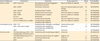
In several early phase trials to date, durable RRs were observed in patients with recurrent EOC and variable PD-1/PD-L1 expression (Table 1) [62636465].
Table 1
Selected trials of PD-1/PD-L1 and CTLA-4 immune checkpoint blockade in ovarian cancer

| Target | Antibody | IgG subclass | Study setting | Phase | No. | CR | PR | SD | ORR (%) | DCR (%) | Median PFS (wk) | ≥G3 AE (%) | Trial identifier | Ref. |
|---|---|---|---|---|---|---|---|---|---|---|---|---|---|---|
| PD-1 | Nivolumab | Human IgG4 | Relapsed platinum resistant EOC | II | 20 | 2 | 1 | 6 | 15.0 | 45.0 | 14.0 | 40.0 | UMIN000005714 | [62] |
| Pembrolizumab | Humanized IgG4 | Advanced EOC | I | 26 | 1 | 2 | 6 | 11.5 | 34.6 | NA | 3.8 | NCT02054806 | [63] | |
| PD-L1 | BMS-936559 | Human IgG4 | Advanced EOC | I | 17 | 0 | 1 | 3 | 6.0 | 23.5 | NA | 9.0 | NCT00729664 | [64] |
| Avelumab | Human IgG1 | Relapsed platinum resistant EOC | I | 124 | 0 | 12 | 55 | 9.7 | 54.0 | 11.3 | 6.5 | NCT 01772004 | [65] | |
| CTLA-4 | Ipilimumab+GM-CSF | Human IgG1 | Advanced EOC | I | 9 | 0 | 1 | 3 | 11.1 | 44.4 | NA | 22.2 | NCT01611558 | [67] |
AE, adverse events; CR, complete response; CTLA-4, cytotoxic T-lymphocyte-associated antigen 4; DCR, disease control rate includes patients with complete response, partial response and stable disease; EOC, epithelial ovarian cancer; GM-CSF, granulocyte-macrophage colony-stimulating factor; IgG, Immunoglobulin G; NA, data not available at the time of review; NCT, National Clinical Trial; ORR, overall response rate; PD-1, programmed cell-death 1; PD-L1, programmed cell-death ligand 1; PFS, progression free survival; PR, partial response; SD, stable disease; UMIN, University Hospital Medical Information Network.
Interestingly, consistent, durable and significant responses were observed when patients with recurrent EOC were treated with checkpoint inhibitors. In a phase II platinum-resistant EOC trial, nivolumab was administered in 2 cohorts. Two patients achieved CR with ORR of 17% and disease control rate (DCR) (CR/partial response [PR]/SD) of 44% observed [62]. 80% of tumor specimen showed high expression of PD-L1 but no significant correlation with response was observed [62]. A study of pembrolizumab in patients with PD-L1 positive advanced solid tumors (PD-L1 expression ≥1%) in a phase Ib trial presented by Varga and colleagues [63] showed 1/26 patients with advanced EOC obtained a CR while 2/26 experienced PR. Best ORR was 11.5% and DCR was 34.6%. BMS-936559 was also used in 207 patients with advanced solid tumors, including 17 EOC patients. One-seventeenth and 3/27 obtained PR and SD respectively [64]. Avelumab was used in 124 women with recurrent and refractory EOC in a phase Ib trial that observed 12 PR and ORR of 10.7% (DCR 54.7%) with grade ≥3 immune related RRs of 6.5% (Table 1) [65]. The durable RRs in a subset of ovarian cancer and ease of tolerability of these checkpoint inhibitors support further investigation, however, it is clear that only a proportion of patients will respond to checkpoint inhibitor monotherapy. Of note, there were a total of 3 reported cases of recurrent OCCC treated in the nivolumab and avelumab studies with durable responses in all 3 patients [6265]. These results are indeed intriguing and further clinical trials are warranted.
3. Clinical studies of CTLA-4 blockade in advanced ovarian cancer
In 2003, the CTLA-4 antibody, ipilimumab, was used in patients with advanced ovarian cancer previously immunized with ovarian tumor cell vaccine transduced with granulocyte-macrophage colony-stimulating factor (GM-CSF). A single infusion of ipilimumab at 3 mg/kg induced a durable stabilization or reduction of cancer antigen 125 (CA-125) levels in 2 out of 2 ovarian cancer patients [66] while in an expanded cohort, SD was seen in 3 patients with 1 patient achieving a durable response lasting more than 4 years [67]. Two patients experienced grade 3 gastrointestinal toxicities and tumor regression correlated with CD8+/regulatory T (Treg) ratio (Table 2) [67]. No clinical evidence for tremelimumab, the other anti-CTLA-4 antibody, is yet available for ovarian cancer.
Table 2
Selected ongoing trials of immune checkpoint inhibitors in gynecological cancers
aCD-27, agonist monoclonal antibody for CD27; aCSF1R, small-molecule receptor tyrosine kinase inhibitor of CSF1R; aCTLA-4, anti-cytotoxic T-lymphocyte-associated antigen 4; aPD-1, anti-programmed cell-death 1; aPD-L1, anti-programmed cell-death ligand 1; CTX, chemotherapy; JAK1i, inhibitor of Janus-associated kinase 1; NCT, National Clinical Trial; PI3Kδi, poly (ADP-ribose) polymerase inhibitor; PI3Kδi, inhibitor of the delta isoform of phosphoinositide-3 kinase; TLRa, agonist of Toll-like receptor 8; VEGFi, inhibitor of vascular endothelial growth factor.
4. Combination blockade and other strategies in advanced ovarian cancer
To date, immune checkpoint inhibitors tested as single agents in the setting of relapsed platinum resistant EOC have shown ORR <15%. In order to generate clinically effective antitumor immune responses, several immune processes may need to be manipulated simultaneously using combinatorial approaches of conventional therapy, novel targeted agents and immunotherapy (Table 3). Preclinical studies of combination immunotherapy in ovarian cancer are encouraging revealing up to half of TILs were positive for both CTLA-4 and PD-1 in the murine model [68]. These TILs had lower proliferative abilities and did not generate the necessary cytokines to mediate cell-kill. However, with dual blockade of both immune checkpoints, TIL function was restored and responses observed were double that of either agent given alone [68]. Clinical trials combining PD-1 inhibitors with chemotherapy, targeted therapy, and other forms of immunotherapy are currently underway. A phase I study of combination ipilimumab with nivolumab in metastatic melanoma showed increased PFS compared with monotherapy treatment [69], thus generating much interest in other solid tumors, including ovarian cancer (NCT02498600). However, 55.0% of patients receiving the combination regimen experienced grade 3 or 4 treatment-related adverse event of which the most common were diarrhea (9.3%) and colitis (7.7%), compared with 16.3% (nivolumab) and 27.3% (ipilimumab) in patients treated in the monotherapy arms [69]. These toxicities will certainly need to be considered in the context of heavily pre-treated ovarian cancer patients.
Table 3
Potential drug combinations with immune checkpoint blockade relevant for gynecological cancers
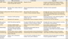
| Drug class | Effect on the tumor | Combination | Rationale | Ref. |
|---|---|---|---|---|
| PARP-inhibitor | Inhibit repair of double strand DNA breaks through PARP inhibition | Olaparib+CTLA-4 | Increase recruitment of TILs through inhibition of DNA repair and induction of memory T cells in BRCA mutant or C2/immune reactive EOC | [52] |
| [53] | ||||
| [54] | ||||
| [70] | ||||
| VEGF inhibitor | Modulate TME to reduce MDSC and Treg cells | Bevacizumab/cediranib+immune checkpoint inhibitor | Eliminate immune suppression | [96] |
| [97] | ||||
| [99] | ||||
| PI3K/AKT pathway inhibitor | Inhibit PI3K-AKT pathway activation | AKT inhibitor/PI3K inhibitor/mTOR inhibitor+immune checkpoint inhibitor | Abrogate anti-tumor immune resistance resulting from PI3K-AKT pathway activation; decreases pro-survival signaling and decreases tumor-promoting inflammation | [84] |
| Metformin | Activation of AMPK pathway | Metformin+immune checkpoint inhibitor±GM-CSF | Restoring exhausted T-cells potentiating effector T-cell and macrophage activity | [93] |
| GM-CSF | Adjuvant to DC maturation | Sargramostim+immune checkpoint inhibitor | Activate de novo immune response | [96] |
| [113] | ||||
| Wnt pathway inhibition | Inhibit Wnt pathway signaling | Porcupine inhibitor/β catenin inhibitor/FZD antibodies+immune checkpoint inhibitor | Abrogate anti-tumor immune resistance resulting from activation of Wnt pathway | [86] |
| [87] | ||||
| HSP90 | Increases unfolded protein-associated stress in tumor cells | HSP90+vaccine±immune checkpoint inhibitor | Boost antigen recognition of tumor cells; decreases antigen presentation | [116] |
| [117] | ||||
| Radiotherapy | Increase antigen presentation; stimulation of danger signaling pathways | Radiotherapy→(sequential) immune checkpoint inhibitor | Eliminate immune suppression; induce immunogenic cancer death and enhance antigen presentation | [110] |
| [111] | ||||
| Conventional chemotherapy | Increase release of tumor antigens and damage-associated molecular pattern molecules | Anthracyclines/platinum/cyclophosphamide chemotherapy+immune checkpoint inhibitor | Upregulate neoantigens; induce immunogenic cancer death and enhance antigen presentation | [96] |
| [106] | ||||
| [107] | ||||
| Dual immune checkpoint blockade | Inhibits T cell co-stimulatory receptor | CTLA-4+PD-1/PD-L1 inhibitor PD-1/PD-L1 inhibitor+LAG-3/TIM-3 inhibitors | Elimination of immune suppression by inhibiting immune suppressive mediators | [100] |
| [102] | ||||
| [103] |
AMPK, AMP-activated protein kinase; AKT, protein kinase B; CTLA-4, cytotoxic T-lymphocyte-associated antigen 4; DC, dendritic cell; EOC, epithelial ovarian cancer; FZD, frizzled; GM-CSF, granulocyte-macrophage colony-stimulating factor; HSP90, heat shock protein 90; LAG-3, lymphocyte-activation gene 3; MDSC, macrophage derived suppressor cells; mTOR, mechanistic target of rapamycin; PARP, poly (ADP-ribose) polymerase; PD-1, programmed cell-death 1; PD-L1, programmed cell-death ligand 1; PI3K, phosphatidylinositol 3-kinase; TIL, tumor infiltrating lymphocyte; TIM-3, T-cell immunoglobulin and mucin protein 3; TME, tumor microenvironment; Treg cells, T regulatory cells; VEGF, vascular endothelial growth factor.
Other awaited studies are investigating the combination of immune checkpoint inhibitors with cytotoxic chemotherapy, PARP-inhibitors or VEGF inhibitors (Table 3). Preclinical data in a murine ovarian cancer model demonstrated CTLA-4 antibody but not the inhibition of PD-1/PD-L1 pathway, synergize with PARP-inhibitors increasing long term survival in the majority of mice [70]. There are several trials underway with tremelimumab+olaparib evaluating the potential efficacy of this combination (NCT02571725, NCT02485990). Nevertheless, preliminary phase I data of the anti-PD-L1 antibody, durvulamab, in combination with the PARP-inhibitor, olaparib or VEGF receptor inhibitor, cediranib has demonstrated that this approach is safe and tolerable [71] with increased efficacy. In essence, facilitating the therapeutic targeting of several described hallmarks of cancer in the same effort [72]. Indeed, 9 patients with advanced EOC or breast cancer were treated with durvulamab and olaparib, 1 patient achieved PR, and 5 SD, resulting in a 67% DCR. Interestingly, all patients were BRCA1/2 wild-type in this group. Similar efficacy trends were observed in 7 evaluable patients with advanced ovarian, cervical cancer and uterine leiomyosarcoma on durvulamab and cediranib (57% DCR; 2 PR; 2 SD) [71].
IMMUNE CHECKPOINT INHIBITORS FOR CERVICAL CANCER
Cervical cancers are unique among gynecologic malignancies due to its well-established infectious etiologic agent, chronic human papillomavirus (HPV) infection [14]. This has led to the development of effective preventative vaccines and cytologic screening and DNA testing of HPV types in developed countries leaving the global burden of cervical cancer disproportionately to developing, resource-poor countries. Despite advances in screening, vaccination, and treatment of early-stage disease, advanced stage cervical cancer remains a principal cause of gynecologic cancer mortality with limited treatment options. The Gynecologic Oncology Group (GOG) 240 trial demonstrated improved survival outcomes with the addition of an anti-VEGF agent, bevacizumab, to standard chemotherapy with no significant deterioration in quality of life [73]. Nonetheless, it is unclear what the optimal treatment options are once patients progress on anti-angiogenic therapy. Novel therapeutic strategies warrant urgent investigation in an effort to further improve outcomes.
1. Rationale for immune checkpoint inhibitors in cervical cancer
Recent evidence supports a potential role for immune checkpoint inhibitors as a viable therapeutic strategy in cervical cancers. PD-L1 expression has been reported in 95% of cervical intraepithelial neoplasia and 80% of squamous cell carcinomas [74] and lymph nodes harboring metastatic cervical cancer were characterized by high levels of PD-L1+APCs and FOXP3+ Treg cells [75]. Recently, the role of the PD-1:PD-L1 interaction in HPV-associated head and neck squamous cell cancer (HPV-HNSCC) creating an “immune-privileged” site for initial viral infection and subsequent adaptive immune resistance once tumors are established suggest a rationale for therapeutic blockade of this pathway in patients with HPV-associated tumors [76].
2. Clinical trials in cervical cancer with immune checkpoint inhibitors
To date, there is limited data on reported cases of recurrent/persistent cervical cancer treated with anti-PD-L1 in the literature. The largest study is a phase Ib study, KEYNOTE-028 (NCT02054806), in patients with advanced cervical squamous cell carcinoma treated with pembrolizumab [77]. All patients had ≥1% PD-L1 expression in tumor or stromal cells. A dose of 10 mg/kg of pembrolizumab appeared to be well tolerated in this series of 24 patients with ORR of 12.5%; median duration of response of 19.3 weeks and 6-month PFS rate of 66.7% [77]. These results appear encouraging and several other cervical cancer-directed trials are currently underway (Table 2).
CHECKPOINT INHIBITORS FOR GYNECOLOGICAL CANCER: FUTURE PERSPECTIVES
As we continue to explore the role of immune checkpoint inhibitors in our therapeutic paradigm for gynecologic cancers, several potential hurdles need to be addressed. Firstly, potential biomarkers to determine which immune checkpoint pathways dominate in a particular tumor will be crucial to guide therapy. Secondly, we need a better understanding of the key immune modulatory mechanisms in gynecologic cancers, particularly the causes of immune resistance, and to develop approaches to enhance the efficacy of checkpoint inhibitors. Both these strategies will be discussed in this section.
1. Predictive biomarkers for immune checkpoint inhibitors
As PD-1 is expressed on a large proportion of TILs [78] and the PD-L1 ligands are commonly upregulated on the tumor cell surface of solid tumors [9], the expression patterns of PD-1 ligands may be crucial for determining the suitability of therapeutic blockade of this pathway. Currently, no pre-treatment biomarker has been validated to be included as part of the standard-of-care decision-making for immune checkpoint inhibitors in gynecological cancers. In ovarian cancer, no correlation was found between PD-L1 expression and anti-tumor response in two trials utilizing nivolumab and avelumab [6265]. This is partly due to a wide array of companion diagnostics being developed individually by several pharmaceutical companies each utilizing different IHC antibody clones, protocols, scoring systems and positive cutoffs in determining expression levels [79]. Furthermore, the lack of a uniform consensus in the level of protein expression of key ligands (PD-L1/PD-L2) or receptor (PD-1) in tumor tissue or TME and the dynamic nature of PD-L1 expression makes determining potential biomarkers challenging [79]. Current evidence suggests that the presence of TIL infiltrates and an active PD-1/PD-L1 axis are important for responses to immune checkpoint inhibitors [808182]. In advanced non-small-cell lung cancer (NSCLC), PD-L1 expression of TILs in the TME, not tumor cells, was associated with response in patients treated with a PD-L1 inhibitor, atezolizumab [82], while reports have correlated TILs expressing PD-L1 at the invasive margins of melanoma cells with response to pembrolizumab [81]. Moreover, in ovarian cancer, tumor associated macrophages (TAMs) in the TME frequently express PD-L1 and may be relevant in response to checkpoint inhibitors [83]. Given that a uniform consensus for predictive biomarkers remains elusive, perhaps the use of surrogate biomarkers could be useful for predicting response. There is potential for specific oncogenic pathways, such as phosphatidylinositol 3-kinase (PI3K)-protein kinase B (AKT) or signal transducer and activator of transcription 3 (STAT3) to induce the expression of specific immune-inhibitory molecules and could thus be used as surrogate markers [184] of immune checkpoint therapy resistance. Established molecular markers in certain tumor types could also potentially be used as predictive biomarkers. As mentioned previously, a strong association between BRCA1 inactivation with the C2/immunoreactive molecular subtype was recently observed [53]. It is still unclear whether BRCA1-mutation associated tumors tend to be more immunogenic. Recent interrogation of the Cancer Genome Atlas (TCGA) dataset observed the well-established prognostic benefit of CD8+ TIL restricted to tumors that additionally harbor plasma cells but no association was observed with BRCA1/2 status. They also found that, rather than working in opposition, the B-cell and T-cell lineages mount closely integrated responses to human tumors as reflected by their physical co-localization, synergistic functional profiles, and interdependent prognostic significance [50]. However, the knowledge of BRCA1/2 mutation status should still be considered in the design of future immunotherapy trials. As previously described, MSI high tumors have been observed to harbor a higher mutational load and expression of neoantigens, which may account for improved responses to immune checkpoint inhibitors [31]. Further validation of the utility of this molecular aberration as a predictive marker for gynecological malignancies will depend on the outcome of ongoing trials.
2. Overcoming immune resistance to enhance effectiveness of immune checkpoint inhibitor
In most tumors, PD-L1 is not constitutively expressed but rather induced on tumor cells by inflammatory signals (i.e., IFN-γ) to resists immune elimination [1]. Evidence suggests that for therapeutic PD-1 blockade to induce tumor regression, the TME is crucial in modulating an anti-tumor immune response [81]. Patients who responded to anti-PD-1 were observed to contain tumor specific CD8+ T cells expressing PD-1 in the TME in close proximity to PD-L1 expressing cells [81] suggesting importance for modulation of the TME to enhance efficacy of checkpoint inhibitors.
Recent literature have cited four mechanisms that require targeting for successful induction of anti-tumor immunity [10]: (1) Elimination of immune suppression by inhibiting immune suppressive mediators; (2) Inducing immunogenic cancer cell death either with radiation/chemotherapy or targeted treatment; (3) Enhancing antigen presentation function via immune adjuvants; and (4) Potentiating effector T-cell and macrophage activity [10], further emphasizing the need for effective combination strategies to enhance the efficacy of immunotherapy (Table 3).
3. Combination with targeted treatment
There is growing evidence that oncogenic pathways in tumors can promote resistance to checkpoint blockade with activation of the PI3K-AKT pathway and/or phosphatase and tensin homolog (PTEN) loss in patients with melanoma [84] while preclinical studies observed oncogenic epidermal growth factor receptor (EGFR) signaling was able to remodel the TME and trigger PD-1 activation as a mechanism of immune escape in EGFR driven lung tumors [85]. These observations indicate that combinatorial strategies of targeted inhibitors and immune checkpoint inhibitors may be able to potentiate the effects of checkpoint blockade (Table 3). More recently, it was observed in melanoma that intrinsic oncogenic pathway mediated through the WNT-β-catenin signaling pathway prevents T-cells from infiltrating tumors [86]. This could potentially be relevant for a cohort of EOCs that are driven by non-canonical Wnt/PCP pathway [87]. Thus, a combination of agents that inactivate the Wnt signaling pathway with anti-PD-1/PD-L1 agents could potentially improve outcomes in gynecologic cancers (Table 3). Several epidemiologic and meta-analysis have consistently shown metformin use was associated with decreased cancer risk and/or reduced cancer mortality [8889]. Reports suggest metformin exerts its anti-tumor effects mainly through AMP-activated protein kinase (AMPK) activation and PI3K-AKT-mechanistic target of rapamycin (mTOR) inhibition [90]. Indeed, preclinical evidence showed synergistic anti-tumor effects of metformin in combination with chemotherapeutic agents like carboplatin [91] and paclitaxel [92]. Recent reports have hinted that the mechanism may be immune-mediated with preclinical evidence suggesting metformin protects CD8+ TILs from exhaustion and eventual apoptosis by restoring the exhausted PD-1-ve T-cell immunoglobulin and mucin protein 3 (TIM-3)+ CD8+ TILs via a shift from central memory T cells to effector memory T cell phenotype [93] thus potentiating the anti-tumor effects of immune checkpoint inhibitors (Table 3).
Recent data showed the addition of a vascular endothelial growth factor A (VEGFA) inhibitor, bevacizumab, in EOC [439495] and cervical cancer [75] improved outcomes in a subgroup of patients. Certainly, VEGFA is best known for its angiogenic effects. However, this cytokine also exerts immunomodulatory effects that block the maturation of DC and promote expansion of myeloid derived suppressor cells (MDSCs) [96]. DC when loaded with myeloma cells containing VEGFA, were found to display lower levels of co-stimulatory molecules and a reduced ability to stimulate T cells compared to unloaded controls [97]. The suppressive effects of VEGFA was abrogated by the addition of bevacizumab [97], and sunitinib, a multi-tyrosine kinase inhibitor that blocks VEGF receptor function [98] resulting in decreased MDSCs and Treg cell population and function. Furthermore, in the B16 melanoma model, a VEGFA antibody combined with adoptive T cell transfer intensified tumor infiltration, reduced tumor growth and prolonged survival compared with either monotherapy alone [99].
4. Combination with other immune checkpoint inhibitors
Co-targeting of CTLA-4 and PD-1 either in combination or sequential in advanced-stage melanoma patients resulted in significantly improved RRs of 53% [100]. The proposed mechanism of synergism is the amplification of T-cells in lymphoid organs and tumor tissue by anti-CTLA-4, whereas PD-1 inhibition overcomes immune suppression in tumor tissues [101]. Similar combination therapies are now being investigated in ovarian cancer (NCT02498600).
Other immune checkpoint receptors have been implicated in the induction of lymphocyte exhaustion, a state in which T or B cells remains alive but are unable to activate an immune response [1]. Blocking of these specific inhibitory receptors could potentially enhance anti-tumor immunity. The lymphocyte-activation gene 3 (LAG-3) and TIM-3 are examples of other inhibitory receptors commonly expressed on exhausted PD-1+ T-cells and targeting these receptors either alone or in combination with other immune checkpoint inhibitors have been shown to enhance anti-tumor activity in preclinical models [102103].
5. Combination with conventional treatment (chemotherapy, radiotherapy)
Radiotherapy and chemotherapy are the treatment backbone in patients with gynecological cancer and have also been shown to modulate anti-tumor immune response [104]. Radiation and chemotherapeutics can lead to upregulation of neoantigens to activate the adaptive immune system and enhance tumor cell immunogenicity [105]. Agents like anthracyclines and oxaliplatin result in the release of tumor antigens and damage-associated molecular pattern molecules from tumor cells to induce immune cell death [106] by polarizing DCs towards a pro-inflammatory phenotype and increase priming towards TH1 anti-tumor T cells and away from Treg cells [96]. Additionally, some chemotherapeutic agents such as cyclophosphamide at low doses down regulate Treg cells and have been observed to provide a more favorable TME for cellular immunotherapy [107]. Indeed, preclinical evidence demonstrates improved efficacy with the combination of radiation and/or chemotherapeutics with immunotherapy, compared with the use of either agent alone [108].
There have been case reports highlighting a phenomenon in which local radiotherapy in combination with immune checkpoint blockade was associated with regression of metastatic cancer at a distance from the irradiated site, known as the abscopal effect [109]. The biologic characteristics underlying this effect is thought to be due to the release of clastogenic factors from irradiated cells into serum inducing chromosomal damage in un-irradiated cells [110]. Furthermore, recent preclinical evidence showed ablative radiation-initiated immune response and tumor reduction were greatly amplified by immunotherapy [110111], with radiation observed to enhance recruitment of TIL and upregulate PD-L1 expression [111]. Recent conflicting evidence has emerged demonstrating increased subclonal (present only in a subset of tumor cells) neoantigens from previous cytotoxic treatment resulted in increased intratumoral heterogeneity and reduce response to checkpoint blockade [112]. Hence, more immunotherapy trials with robust translational framework will be required to further explore these observations while taking into account their inherent toxicities.
6. Combination with vaccines
Preclinical studies have observed dramatic synergy between tumor vaccines and the immune checkpoint inhibitors previously described in poorly immunogenic models [113114]. Multiple vaccine interventions that activate de novo immune response may not appear to induce tumor regression because tumors up regulate immune checkpoint ligands as an escape mechanism [1]. Hence combining these two approaches may induce increase efficacy in patients that would not have responded to either therapy alone. GM-CSF is a potent immune adjuvant to DC maturation used in several tumor vaccine trials. When combined with CTLA-4 blockade, this vaccine combination was found to increase inflammatory infiltrates and tumor regression [6667], suggesting that vaccine-induced anti-tumor T cells were present within the tumor but energized owing to CTLA-4 co-inhibition. Similarly, in a preclinical study combining vaccines and PD-1 blockade, mice receiving combination therapy had increased overall survival and decreased tumor growth compared to either therapy alone [113] proving that synergistic mechanisms exist with bolstering tumor antigen presentation and T cell priming with checkpoint immune blockade.
CONCLUSION
Promising preclinical and clinical data supports the further development of immune checkpoint inhibitors in gynecologic cancers. However, despite encouraging clinical outcomes, only a proportion of patients benefit from this treatment. As such, concerted efforts to define biomarkers that can predict the groups of patients and the type of cancer that will respond to immune checkpoint blockade are urgently required. The challenges faced reflects the complex interplay of host immune-cell populations within tumor and TME which is further complicated by intra- and inter-tumoral heterogeneity [115]. As the era of immunotherapy dawns on the field of oncology, the ability to fully elucidate this complex network will be the crucial step in facilitating a personalized approach to target tumors on several fronts, and ultimately improving patient outcomes.
 XML Download