

 PDF
PDF ePub
ePub Citation
 Print
Print


INTRODUCTION
According to the Barcelona Clinic Liver Cancer staging, extrahepatic metastasis of hepatocellular carcinoma (HCC) is a poor prognostic factor, and is regarded as advanced stage (C). Most patients with advanced stage receive palliative treatment. The most common site for extrahepatic metastasis of HCC is the lung, followed by regional lymph node, and bone [1,2]. Metastatic skin lesion of HCC is rare, accounting for less than 0.8% of all metastatic cutaneous tumors and occurring in 2.7%-3.4% of all HCCs [3,4]. Skin metastasis is a sign of poor prognosis, indicating the strong possibility of metastases in other regions of the body, and points to a median survival time of less than five months [5,6]. We report on a case of multiple metastases, including skin metastasis, which were treated with multidisciplinary therapy.
CASE REPORT
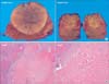
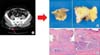
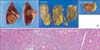
A 52-year-old male patient with a history of type B viral hepatitis visited the hospital with abdominal distension; at that time, a huge mass (a mass measuring approximately 7 cm on segment 8, and a mass measuring 2.5 cm on segment 6) in the right hemiliver and hematomas around the liver were observed on abdominal computed tomography (CT) scan; the diagnosis was HCC rupture (Fig. 1). After transarterial chemoembolization (TACE), segment 8 segmentectomy and segment 6 wedge resection were performed (Fig. 2); hyperthermic intraperitoneal adriamycin irrigation was then performed for prevention of peritoneal seeding. According to the biopsy report, there was no viable tumor because of TACE. The patient was discharged with no specific complication and no evidence of recurrence was observed for seven years postoperative (During the seven years, ultrasonography or CT and tumor marker check was performed every 3 months, but there was no abnormal finding). However, 84 months after the initial operation, an abnormal splenic mass was observed on follow-up ultrasonography, and his tumor marker was elevated (α-fetoprotein, 1.94; protein induced by vitamin K absence or antagonist II, 309). Therefore, we performed positron emission tomography-CT, and multiple metastases were observed. The sites of metastases were brain, adrenal gland, spleen, kidney, skin (buttock), back muscle, and buttock muscle (Figs. 3, 4); however, there was no intrahepatic recurrence. Skin biopsy confirmed metastasis of HCC (Fig. 5). The patient then received radiation therapy for brain metastasis, and took oral medication with sorafenib (Nexavar, Bayer Health Care, Leverkusen, Germany). After one month, a new lesion was observed on his right thumb (Fig. 6). However, follow-up abdominal CT showed stable condition of the previously mentioned multiple metastases (mass of spleen, left adrenal gland, left kidney, back muscle, and buttock muscle), and the brain mass had shrunk. We performed en bloc resection of intra-abdominal metastatic masses (splenectomy, left adrenalectomy, left nephrectomy) and excision of the right thumb mass (Fig. 7; Edmonson-Steiner grade of these masses are grade 2, and histologic type is trabecular, and cell type is hepatic cell). Currently, 18 months after initial diagnosis of multiple metastases, he is still alive, and he takes an oral medication with sorafenib for target therapy of remnant tumor in back muscle, buttock, and brain, which are unresectable (Fig. 8).
DISCUSSION
Metastatic skin lesion of HCC is rare; fewer than 50 such cases have been reported in the literature with most of them diagnosed histologically by excision biopsy specimen [7]. Cutaneous metastases originating from HCC appear as rather rapidly growing nodules, usually on the scalp, chest, and shoulder. They appear as single or multiple, firm, painless, nonulcerative, reddish-blue nodules, measuring 1 to 2.5 cm in diameter [8]. Especially in cases of skin metastasis of HCC caused by systemic spread of disease, indicating the strong possibility of metastases in other regions of the body, the prognosis is dismal [5,6]. In addition, in this case, the patient had multiple metastatic lesions, including skin metastasis. Despite these poor prognostic factors, this patient seemed to have good tumor biology, for example, 100% tumor necrosis after initial TACE, recurrence after long period, good response to treatment after multiple site recurrence, and not highly elevated AFP. This favorable tumor biology may be an important factor of long-term survival in this patient.
In treatment of advanced HCC, to date, the only drug that has successfully made it into a phase III trial is sorafenib. Sorafenib is an oral multikinase inhibitor blocking tumor cell proliferation and angiogenesis by targeting the serine/threonine kinases Raf-1/B-Raf and the tyrosine kinases of vascular endothelial growth factor receptor-2/-3 and Platelet-derived growth factor receptors-b. Median survival and the time to radiologic progression were nearly three months longer for patients treated with sorafenib than for those who received placebo [9]. In this case, after diagnosis of multiple metastases, he was treated with sorafenib. Although we performed successful resection of intraabdominal metastatic masses, remnant tumors remain (brain, back muscle, and buttock muscle). Therefore, further survival of this patient is mainly dependent on sorafenib.


 XML Download