Journal List > Ann Lab Med > v.37(1) > 1091574

Choi, Jang, Jeong, Jeon, Lee, Shin, Hong, and Lee: A Case of Chronic Myeloid Leukemia With Rare Variant ETV6/ABL1 Rearrangement
Dear Editor,
The translocation (9;12)(q34;p13) ETV6/ABL1 rearrangement is a rare but recurrent chromosomal translocation associated with a variety of hematological malignancies, including CML, atypical CML, AML, and ALL [1]. The structure of the ETV6/ABL1 oncoprotein is similar to that of BCR/ABL1, and they initiate similar downstream pathways [2]. There are two ETV6/ABL1 fusion isoforms: the type A isoform, which fuses ETV6 exon 4 with ABL1 exon 2; and the type B isoform, which fuses ETV6 exon 5 with ABL1 exon 2 [3, 4]. To date, 30 cases of ETV6/ABL1 fusion have been reported [5, 6], and only one of these cases resulted in CML with positive BCR/ABL1 rearrangement [7]. Herein, we report a rare case of CML with ETV6/ABL1 rearrangement.
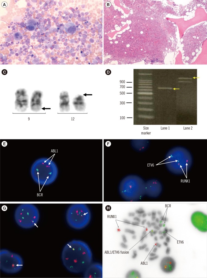
A 54-yr-old male was admitted with persistent leukocytosis. Complete blood counts showed a white blood cell count of 21.7×10⁹/L with 1% blasts, Hb of 126 g/L, and platelet count of 294×109/L. Physical examination was unremarkable. Bone marrow (BM) analysis showed typical characteristics of CML (Fig. 1A, B). Chromosomal analysis of the BM cells demonstrated a balanced t(9;12)(q34;p13) translocation, which was not the Philadelphia chromosome (Fig. 1C). FISH analysis with probes for BCR/ABL1 (Abbott Vysis, Des Plaines, IL, USA detected no fusion signal. However, reverse transcriptase (RT)-PCR analysis of the BCR/ABL1 fusion transcripts yielded positive results; the reaction product was 700 bp long, indicating positive rearrangement and hence, presence of the P230 chimeric protein at the molecular level (Fig. 1D).
To visualize the ETV6/ABL1 fusion signal, we prepared a mixture of two commercially available, locus-specific identifiers: a BCR/ABL1 dual color, dual fusion translocation probe, and an ETV6/RUNX1 extra signal dual color translocation probe (Abbott Vysis) (Fig. 1E, F). Metaphase and interphase FISH with the mixed BCR/ABL1 and ETV6/RUNX1 probes showed one yellow fusion signal at 9q34, which was derived from a green signal from ETV6 and a red signal from ABL1 (Fig. 1G, H). RT-PCR analysis of the ETV6/ABL1 fusion transcript was positive for the 1,141-bp product, indicating a type B fusion (Fig. 1D). After diagnosis, the patient was transferred to another hospital, and therefore, follow-up BM examination was not possible.
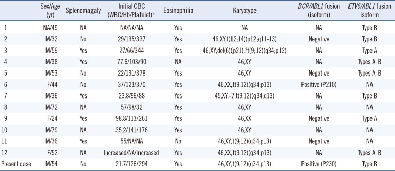
ETV6/ABL1 rearrangement has been reported to result in enhanced tyrosine kinase activity and neoplastic transformation [3, 8]. A total of 13 cases of ETV6/ABL1-positive or atypical CML have been reported to date (Table 1) [5, 7]. Among those cases, including the present case, two were BCR/ABL1 fusion-positive and 11 were either unknown or negative for the BCR/ABL1 fusion. Both BCR/ABL1 fusion-positive cases presented with persistent leukocytosis, eosinophilia, and no splenomegaly. Their pathological findings were consistent with those for CML, but BCR/ABL1 rearrangement was not confirmed by karyotyping or FISH. Only RT-PCR revealed the rearrangement, and the amplicon size was 504 bp [7] and 700 bp in the present case, respectively. Marked eosinophilia, which is a common characteristic of the ETV6/ABL1 translocation [7], was also predominant. Although the pathogenesis of eosinophilia is not clearly understood, ETV6 is known to play an active role in the commitment of hematopoietic myeloid precursors to eosinophilic differentiation [9].
Rare cases of CML are associated with a BCR breakpoint that is considerably more directed towards the 3' end than the major breakpoint cluster region, which encodes a P230 BCR/ABL1 fusion protein. Our patient had a novel-sized BCR/ABL1 fusion transcript (700 bp), which is ~140 bp smaller than the typically observed micro BCR/ABL1 (c3a3) amplicon size of 838 bp, suggesting in-frame deletion of an exon. Although the lack of the Philadelphia chromosome observed by karyotyping and FISH is unusual, it is possible that RT-PCR is more sensitive than cytogenetics or FISH. Unfortunately, Sanger sequencing of the identified novel transcript could not be performed, and 2 weeks later, a repeat RT-PCR analysis of the peripheral blood failed to detect any BCR/ABL1 fusion transcripts.
Detection of the ETV6/ABL1 fusion may help to inform treatment plans for patients with rare hematologic malignancies. In these cases, tyrosine kinase inhibitors can be effective because of the significant overlap between the molecular targets of ETV6/ABL1 and those of BCR/ABL1 [10].
In conclusion, we identified an ETV6/ABL1 translocation in a patient with CML, which was confirmed by FISH with combined BCR/ABL1 and ETV6/RUNX1 probes, as well as by RT-PCR analysis. This report will contribute to a better understanding of the clinical phenotype and molecular basis of this rare type of ETV6/ABL1-positive hematologic malignancy.

Acknowledgments

This work was supported by the Soonchunhyang University Research Fund.

Notes

Authors' Disclosures of Potential Conflicts of Interest: No potential conflicts of interest relevant to this article were reported.

References

1. De Braekeleer E, Douet-Guilbert N, Morel F, Le Bris MJ, Basinko A, De Braekeleer M. ETV6 fusion genes in hematological malignancies: a review. Leuk Res. 2012; 36:945–961. PMID: 22578774.
2. Malinge S, Monni R, Bernard O, Penard-Lacronique V. Activation of the NF-kappaB pathway by the leukemogenic TEL-Jak2 and TEL-Abl fusion proteins leads to the accumulation of antiapoptotic IAP proteins and involves IKKalpha. Oncogene. 2006; 25:3589–3597. PMID: 16434962.
3. Park J, Kim M, Lim J, Kim Y, Han K, Kim JS, et al. Variant of ETV6/ABL1 gene is associated with leukemia phenotype. Acta Haematol. 2013; 129:78–82. PMID: 23171811.
4. Million RP, Harakawa N, Roumiantsev S, Varticovski L, Van Etten RA. A direct binding site for Grb2 contributes to transformation and leukemogenesis by the Tel-Abl (ETV6-Abl) tyrosine kinase. Mol Cell Biol. 2004; 24:4685–4695. PMID: 15143164.
5. Gancheva K, Virchis A, Howard-Reeves J, Cross NC, Brazma D, Grace C, et al. Myeloproliferative neoplasm with ETV6-ABL1 fusion: a case report and literature review. Mol Cytogenet. 2013; 6:39. PMID: 24053143.
6. Song JS, Shin SY, Lee ST, Kim HJ, Kim SH. A cryptic ETV6/ABL1 rearrangement represents a unique fluorescence in situ hybridization signal pattern in a patient with B acute lymphoblastic leukemia. Ann Lab Med. 2014; 34:475–477. PMID: 25368826.
7. Keung YK, Beaty M, Steward W, Jackle B, Pettnati M. Chronic myelocytic leukemia with eosinophilia, t(9;12)(q34;p13), and ETV6-ABL gene rearrangement: case report and review of the literature. Cancer Genet Cytogenet. 2002; 138:139–142. PMID: 12505259.
8. De Braekeleer E, Douet-Guilbert N, Rowe D, Bown N, Morel F, Berthou C, et al. ABL1 fusion genes in hematological malignancies: a review. Eur J Haematol. 2011; 86:361–371. PMID: 21435002.
9. La Starza R, Trubia M, Testoni N, Ottaviani E, Belloni E, Crescenzi B, et al. Clonal eosinophils are a morphologic hallmark of ETV6/ABL1 positive acute myeloid leukemia. Haematologica. 2002; 87:789–794. PMID: 12161353.
10. Perna F, Abdel-Wahab O, Levine RL, Jhanwar SC, Imada K, Nimer SD. ETV6-ABL1-positive "chronic myeloid leukemia": clinical and molecular response to tyrosine kinase inhibition. Haematologica. 2011; 96:342–343. PMID: 21193423.
Fig. 1

Bone marrow (BM) aspiration, biopsy, karyotyping, reverse transcription (RT)-PCR, and FISH analyses of the present case. (A) BM aspiration (Wright-Giemsa stain, ×400) and (B) BM biopsy (hematoxylin & eosin stain, ×50) revealed 90% hypercellular marrow with a left-shifted neutrophilic series, an increased number of eosinophilic precursors, and small, hypolobated megakaryocytes. (C) Karyotyping showing t(9;12)(q34;p13); arrows indicate the translocated regions. (D) RT-PCR using BCR/ABL1 and ETV6/ABL1 primer pairs, revealing the 700-bp BCR/ABL1 (lane 1, arrow) and 1,141-bp ETV6/ABL1 (lane 2, arrow) fusion transcripts; left, 100-bp molecular weight marker ladder. The internal control 911-bp band is shown in lane 2. (E and F) FISH analysis using BCR/ABL1 or ETV6/RUNX1 probes, showing no abnormal signal. (G and H) FISH using the mixed BCR/ABL1 and ETV6/RUNX1 probes revealing one yellow fusion signal (ABL1, red; ETV6, green) on 9q34 analyzed in interphase (G) and metaphase (H) cells.

alm-37-77-g001
Table 1

Summary of patients with CML or atypical CML carrying the ETV6/ABL1 fusion transcript

alm-37-77-i001
Sex/Age (yr) Splenomagaly Initial CBC (WBC/Hb/Platelet)* Eosinophilia Karyotype BCR/ABL1 fusion (isoform) ETV6/ABL1 fusion isoform
1 NA/49 NA NA/NA/NA Yes NA NA Type B
2 M/32 No 29/135/337 Yes 46,XY,t(12;14)(p12;q11–13) Negative Type B
3 M/59 Yes 27/66/344 Yes 46,XY,del(6)(p21),?t(9;12)(q34;p12) NA Type A
4 M/38 Yes 77.6/103/90 NA 46,XY NA Types A, B
5 M/53 No 22/131/378 Yes 46,XY Negative Types A, B
6 F/44 No 37/123/370 Yes 46,XX,t(9;12)(q34;p13) Positive (P210) NA
7 M/36 Yes 23.8/96/88 Yes 45,XY,–7,t(9;12)(q34;q13) NA Type B
8 M/72 NA 57/98/32 Yes 46,XY NA NA
9 F/24 Yes 98.8/113/261 Yes 46,XX Negative Type A
10 M/79 NA 35.2/141/176 Yes 46,XY NA NA
11 M/36 Yes 55/NA/NA No 46,XY,t(9;12)(q34;p13) Negative NA
12 F/52 NA Increased/NA/Increased Yes 46,XX,t(9;12)(q34;p13) NA Types A, B
Present case M/54 No 21.7/126/294 Yes 46,XY,t(9;12)(q34;p13) Positive (P230) Type B

Published cases were reviewed by Gancheva et al [5].

*Values are presented in the International System of Units (WBC, ×109/L; Hb, g/L; Platelet, ×109/L).

Abbreviations: CBC, complete blood count; WBC, white blood cell; F, female; M, male; NA, not available; RT-PCR, reverse transcription PCR.

TOOLS
Similar articles