

 PDF
PDF ePub
ePub Citation
 Print
Print


INTRODUCTION
Osteoarthritis (OA) is the most common form of arthritis, and the leading cause of chronic disability. Many studies showed that the change involving the mechanical property of subchondral bone is the main cause of the OA.[12] Recently, not only mechanical property changes of the bone matrix in the osteoporotic bones, but also chemical changes has been reported.[3] The bone matrix is consist of many protein-rich vibrational modes, corresponding predominantly to collagen features such as amide backbone, protein secondary structure, and side chain composition.[4] These chemical properties provide another source of contrast to bone matrix composition. Therefore, the author hypothesized that chemical composition also change in subchondral matrix of OA knee.
Raman spectroscopy was used to investigate chemical compositional change within the cortices of individual bones. [123] Recently it has been used to investigate chemical composition in the subchondral bone, but there are few researches with human OA femoral condyle.
Therefore, the authors aimed to investigate the differences of chemical composition between subchondral bone of femoral condyle with OA and without OA using Raman spectroscopy.
METHODS
1. Specimen and raman spectroscopy
Specimens were obtained from 10 bones of 10 women cadavers with OA (mean age, 63 years; range, 50 to 76). As controls, the distal femur without OA was obtained from 10 bones of age and gender matched cadavers consisting of 10 women (mean age, 61 years; range, 57 to 65). In OA femur specimen, visible inspection showed the typical features of advanced OA. These feature included erosion of cartilage down to exposed subchondral bone, osteophytes. Control femur specimen without OA had no macroscopic visual pathology or history of musculoskeletal disease, and they had intact cartilage surface.
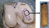
A 10 mm inner diameter cylindrical saw was used to acquire trabecular bone core. Subchondral bone of control group was obtained from medial femoral condyle that was "Central" on sagittal plane and "Central" on frontal plane by International Cartilage Repair Society (ICRS) on cadaver.[4] Subchondral bone of OA group also was obtained from a same region of ICRS grade 3 or 4 cartilage defect on the medial condyle of femur (Fig. 1). The cancellous bone was collected with diameter of 10.0 mm and length of 10 mm, and we further cut 2.5 mm below the subchondral bone plate to avoid cortical plate.
In this study, Raman spectroscopy was employed to determine the chemical information of the cored trabecular bones in the human distal femurs.[56] Surface Enhanced Raman Spectroscopy (SERS) spectra were recorded with a SENTERRA confocal Raman system (Bruker Optics Inc., Billerica, MA, USA). A 785 nm diode laser with 10 mW power at the laser source was used for excitation. The diameter of the focal spot was 2.4 µm with a 20×objective lens (numerical aperture [NA]=0.4). A 10×objective lens (NA=0.25) was used to focus the incident polarized light onto the dimer array. Spectra were recorded over the range of 150-1,800 cm-1 with spectral resolution of 3 cm-1 and integration time of 30s. Baseline correction was performed by the rubber band correction (RBC) with 15 iterations before integrals were calculated to remove the baseline using OPUS 6.5 software (Bruker Optics Ltd, Ettlingen, Germany). The curve-fitting procedure generally yielded bands that were readily identified based on Raman band assignments from previous experiments.
The degree of bone mineral components (phosphate group, carbonate group) and organic components (amide I, amide III, and C-H group) were estimated. The value of the alpha helix to random coil band area ratio was used for collagen quality parameter, which indicates the collagen's secondary structure and the degree of distortion of collagen crosslinks in bone matrix.
RESULTS
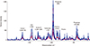
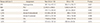
The wavenumber showed a similar trend in the OA and non-OA knees, but Raman intensity showed substantial difference (Fig. 2). The maximum intensity of the phosphate peak (959 cm-1) was 2,376.51±954.6 for the non-OA group and 1,936.3±831.75 for the OA group. The maximum intensity of the phosphate peak (959 cm-1) between the two groups was significantly different (P=0.017). The maximum intensity of the amide I peak (1,668 cm-1) were 474.17±253.42 for the non-OA group and 261.9±205.61 for the OA group. The maximum intensity of the amide I peak were significantly different between the two groups (P=0.042). Also, among other chemical and matrix components (Hydroxyproline, Carbonate, Amide III-disordered; ordered, and CH2), the spectrums showed similar significant differences in the intensity (Table 1).
The value of the alpha helix to random coil band area (alpha helix 1,270 cm-1/random coil 1,240 cm-1) are significantly different (P=0.021), and result showing that there was a trend toward higher collagen maturity for the non-OA group.
DISCUSSION
Most studies regarding the relationship between the condition of normal bones and OA have been focused on the mechanical properties of the subchondral bones. Few studies have reported the changes in mechanical properties and micro-structures of the subchondral bones in early OA.[4567] Moreover, Matsui et al.[8] reported that change of the underlying subchondral bone precedes the degeneration of the joint cartilage. Studies on the matrix of the bones have been carried out mostly in the osteoporosis than OA, and have reported that the bone material properties differed between osteoporotic and normal control groups. In particular, the altered collagen properties at sites of active bone formation explain that affected individuals have osteoblast dysfunction that may play a role in bone fragility.[91011] There were a few published results that shows the difference in the collagen quality between OA and normal group in femoral head or proximal tibia.[1213]
In our study, the values of the alpha helix to random coil band area ratio are also significantly different in subchondral bone from OA group and control group, which reveal that collagen matrix is less ordered in the OA group and the degree of alpha helical structure to disordered structure transition increase with OA group.
But, our study has the limits and considerations for interpreting this result. First, our OA specimens were obtained from region of ICRS grade 3 or 4 cartilage defect on the medial condyle of femur. These results showed just difference between advanced OA group and normal group, and further studies need to investigate the difference of chemical composition according to OA grade. If the chemical component shows difference in earlier OA stage, Raman spectroscopy will be useful for detecting of early OA.
While studies regarding the differences of the mineral to matrix ratio were substantially done, studies figured out detailed differences in the components of chemical composition were rare.[3814] Dehring et al.[15] used murine distal femurs to study the mineral to matrix ratio of the subchondral bone according to the differences in the conditions of cartilage, and reported the usefulness of the Raman spectroscopy. Extracellular matrix of bone is consisted with many chemical properties. The markers of organic matrix have included the amide I band at 1,667 cm-1, the area under the amide I envelope (1,616 to 1,720 cm-1), and CH2 wag at 1,450 cm-1. In this study, 1,450 cm-1 band was used as a marker for the organic matrix because it is less susceptible to conformational changes and thus, it is a more robust measurement of matrix content.[316] We analyzed 7 markers of the properties of subchondral bone and the results showed significant difference between groups. These results lend weight to our hypothesis than the results, which analyzed single chemical component.
The authors used the Raman spectroscopy to figure out the differences in the chemical composition of the subchondral bones. Raman spectroscopy is a spectroscopic technique used to observe vibrational, rotational, and other low-frequency modes in a system. Light from the illuminated spot is collected with a lens and sent through a monochromator. Recently, Raman spectroscopy has been used to evaluate alterations to bone composition associated with osteoporosis. It is believed that the method will be of benefit to the diagnosis and treatment of numerous diseases.[12131617]
Author reported significant changes in the microstructural and mechanical properties in the medial femoral condyle in advanced OA previously.[11] In this study, because the mechanical properties were not measured concurrently, the relationship between the mechanical properties and chemical components cannot be concluded. So, further study needs to investigate how the change of chemical composition affects the mechanical property in the subchondral bone.
 XML Download