

 PDF
PDF ePub
ePub Citation
 Print
Print


INTRODUCTION
Endovascular coil embolization has been an effective treatment modality for the treatment of unruptured intracranial aneurysms. However, major procedural complications have occasionally occurred, including thromboembolism and aneurysm perforation. In addition, vasospasm, hemodynamic ischemia, migration or reconfiguration of coils and subsequent enlargement of the neck or sac were recognized as pivotal limitations of endovascular coil embolization.8) Thromboembolic events are the most common complication of endovascular treatment, and they may be caused by thrombus formation from the catheter or guidewire, or breakdown of the thrombus from the aneurysm, in which coils have been packed, into the parent artery.5) Most thromboembolic complications occur within 48 hours of endovascular treatment; therefore, antiplatelet or anticoagulant is used during the procedure.1) However, delayed thromboembolic event beyond 2 days after coil embolization may occur despite its rarity.
Here, we present a case of delayed symptomatic thromboembolism which occurred 19 days after the coil embolization of an unruptured aneurysm despite antiplatelet therapy.
CASE REPORT
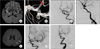
A 56-year-old woman presented to the emergency room with mild left leg numbness of sudden onset. The symptoms had a rapidly improving course over a period of one week. She had hypertension and diabetes mellitus as stroke risk factors. In addition, she had experienced hypertensive intracerebral hemorrhage in the left thalamus five years earlier. She had regularly received antihypertensive medications, oral hypoglycemic agents and aspirin during the past one year. On initial neurological examination, she was alert and had no dysarthria, facial palsy, or language dysfunction. A motor function test revealed mild left leg weakness (MRC grade IV). A tingling sense and numbness in the left hand below the wrist were observed. Deep tendon reflexes and cerebellar function tests were also normal. Her initial National Institutes of Health Stroke Scale (NIIHSS) score was one. The result of her chest x-ray and electrocardiogram showed a normal configuration. A brain magnetic resonance imaging (MRI) scan taken 3 hours 46 minutes after the onset and diffusion-weighted imaging (DWI) showed an acute lacunar infarct in the right thalamus, which may be correlated with small vessel disease, without perfusion defects (Fig 1-A). Antiplatelet agents with 75 mg/day of clopidogrel and 200 mg/day of cilostazol were administered for prevention of recurrent ischemic attacks. A brain magnetic resonance angiogram (MRA) showed a saccular aneurysm which was incorporated with the vessel in the left middle cerebral artery (MCA) bifurcation. A diagnostic transfemoral angiography (TFCA) revealed a 6.4 × 6.0 mm aneurysm with a wide neck (4.2 mm) located at the left MCA bifurcation in the antero-inferior direction (Fig 1-B). Coiling rather than clipping was considered because of acute thalamic infarction, patient's age, and preponderance of the patient. Because of no recurrent ischemic attack or neurologic deficits, treatment timing was considered beyond acute period (minimum 2 weeks after initial attack). Therefore, coil embolization using a multiple catheter technique was performed at hospital day 15. The partial embolization was performed without difficulty because dense packing of the aneurysm was intentionally avoided to prevent protrusion of the Guglielmi Detachable Coils into the parent vessels or occlusion of the parent artery. Consequently, post-procedual imaging could detect some contrast filling in the aneurysmal sac (Fig 1-C, D). The patient was discharged without recurrent or residual neurological deficits and maintained dual antiplatelet agents.
After 14 days of coil embolization, clopidogrel was stopped and 200 mg/day cilostazol was maintained because the patient had no recurrent neurological symptoms. After five more days of single antiplatelet therapy, she visited the emergency room 6 hours after the initial ictus of a language disturbance and right hand clumsiness. A neurological examination showed sensory dominant mixed type aphasia and right arm weakness (MRC grade IV). DWI revealed acute infarction in the territory of the inferior division of the left MCA which was the location of the distal part of the coil embolization (Fig 1-E), and then TFCA was performed without additional images and showed a filling defect as a thrombus in the proximal MCA just distal to the aneurysmal neck (Fig 1-F). Therefore, we performed intra-arterial thrombolytic therapy using 100,000 units of urokinase (for thrombolysis) and 500 mcg abciximab (for inhibiting platelet aggregation) without intravenous tissue plasminogen activator. Immediate post-thrombolysis angiography showed a resolved thrombus and good blood flow to the left MCA branches (Fig 1-G). Language disturbance and motor weakness were not observed. Seven days after the thrombolysis, TFCA revealed still complete recanalization of the thrombosed parent artery. Dual antiplatelet agents were maintained, and she had no recurrent neurological symptoms 12 month after the intra-arterial thrombolytic therapy.
DISCUSSION
We reported a case of delayed thromboembolic complications after endovascular coil embolization of an unruptured MCA aneurysm. In addition, our case had delayed thromboembolic events despite antiplatelet medication.
The peri-procedural ischemic stroke following endovascular coil embolization of aneurysms has been reported in 1 to 28% of cases. Studies with immediate DWI after embolization showed a higher frequency of silent embolism than symptomatic infarct.5)6) These thromboembolic events are widely understood to occur mainly at the time of treatment or within 48 hours of the procedure.1) Most of these strokes can be attributed to thrombosis of the parent or branch arteries from which the aneurysm arises or to distal embolization of the thrombus from the treated aneurysm. The following are possible mechanisms of thrombus formation in coil embolization: 1) anatomical factors (unfavorable configuration, wide neck or low dome-to-neck ratio), 2) procedural factors (multiple microcatheter technique, long procedural time, large volume, long length, or partial embolization), 3) coil factors (coil protrusion or reconfiguration), 4) hemodynamic disturbance, or 5) patient factors (resistant to antiplatelet agent or vulnerable to ischemic insults), or 6) a combination of these factors. Therefore, anticoagulants or antiplatelet agents were used during the period of the endovascular procedure for the prevention of thromboembolic complications. In addition, some delayed thromboembolic events have been reported to occur several days after coil embolization. They were caused by having a large neck,7) coil factors such as prolapse,1) protrusion, or reconfiguration of coils.2)4) In our case, hemodynamic change as stagnant blood flows induced by partial coil embolization of the aneurysm sac and or the patient factor of resistance to antiplatelet medication may theoretically contribute to propagation of thrombus into parent artery, resulting in embolism. However, we did not evaluate patients' response to the antiplatelet drug; thus, this is a limitation of our report.
Two clinical studies reported that antiplatelet preparation during the peri-procedural period reduced thromboembolic complications of elective coil embolization in unruptured aneurysms.3)9) Oral antiplatelet therapy was significantly effective in the reduction of the thromboembolic rate, especially in patients treated by the multiple microcatheter technique.3) Nevertheless, they were focused on the periprocedural period rather than the period afterward. Besides, it has not been determined whether dual antiplatelet therapy is more effective than a single antiplatelet agent on thromboembolic events.
Our patient had a high risk of thromboembolism in that she had an unfavorable aneurysm which led to using multiple microcatheters; moreover partial embolization was performed. Accordingly, in patients with high risk of antiplatelet resistance or ischemic stroke, we postulate that single antiplatelet therapy might be insufficient to prevent a thromboembolic event.
 XML Download