

 PDF
PDF ePub
ePub Citation
 Print
Print


The association between venous thrombosis and malignancy was first described by Trousseau [1] in 1865 and this has been confirmed by several clinical, pathologic and laboratory studies. The thromboembolic event occurs before the diagnosis of cancer, and it has an increased risk of ovarian cancer during the first year of follow-up [2]. A previous report suggests that venous thromboembolism (VTE) may occur as a consequence of underlying cancer, and VTE can be detected prior to the diagnosis of cancer. Some authors estimate that as many as 15% of patients with ovarian cancer will have a thromboembolism [3]. A study on incidental pulmonary embolism (PE) in patients with cancer revealed that the highest prevalence occurred in patients with gynecologic malignancies [4].
We report a case of a 39-year-old patient with ovarian cancer and PE as the initial symptom.
Case Report
A 39-year-old woman presented to the emergency room with the chief complaints of dyspnea and right chest pain that had begun a few days earlier. She was healthy at birth and had no past history of underlying disease or clotting abnormality. There was no history of oral medication, pregnancy, or any other gynecological/surgical problem. On presentation, she was mild febrile, had tachycardia with a regular rate, and her lungs were clear on auscultation. Laboratory studies revealed a hemoglobin value of 10.9 g/dL; prothrombin time 16.5 sec (normal range [NR], 11.0 to 15.0 sec); partial thromboplastin time 45.0 sec (NR, 29.0 to 44.0 sec); D-dimer 40.4 µg/mL (NR<0.4 µg/mL); and C-reactive protein (CRP) 11.53 mg/dL (NR<0.3 mg/dL).
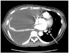
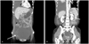
Chest CT revealed pulmonary artery thromboembolism of the left lobe and a massive right pleural effusion (Fig. 1). Abdomino-pelvic CT revealed a 15×12×14 cm solid and cystic mass in the pelvic cavity suggesting ovarian cancer (Fig. 2A) and a wedge-shaped low attenuated lesion in the mid-pole of the left kidney suggesting kidney infarction (Fig. 2B). A cytological examination of the pleural effusion and pleural biopsy revealed metastatic adenocarcinoma. The serum CA-125 level was 768.0 U/mL (NR<35.0 U/mL). Based on these findings; she was diagnosed with ovarian cancer with PE and kidney infarction. Unfractionated heparin (10,000 U/day) was administered as perioperative anticoagulant treatment.
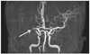
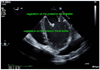
She underwent cytoreductive surgery including total abdominal hysterectomy and bilateral salpingo-oophorectomy. The pathological findings revealed a malignant mullerian mixed tumor of the ovary. After the operation, she complained of swelling and weakness of the left leg. 3D-Angio CT of the lower extremity revealed deep vein thrombosis in the bilateral popliteal and calf veins. An infrarenal inferior vena cava filter was inserted. Brain MR Angiography revealed total obstruction of the right proximal middle cerebral artery (Fig. 3). Trans-esophageal echocardiogram demonstrated some vegetation and mobile echodensity on the atrial surface of the mitral valve (Fig. 4). Infectious endocarditis was suspected with brain infarctions and deep vein thrombosis thus empiric therapy with intravenous antibiotics was initiated. Despite persistent fever and leukocytosis, serial blood cultures remained sterile. Nonbacterial thrombotic endocarditis (NBTE) was suspected. The patient received six cycles of postoperative adjuvant therapy with carboplatin and paclitaxel. Follow-up echocardiograms have shown a gradual regression in the size and the extent of the vegetations. However, her metastatic lung lesion worsened and she died 14 months after her initial visit.
Discussion
PE as a paraneoplastic feature can be caused by a wide range of malignancies (e.g., those of the pancreas, lung, stomach, liver and ovary). Malignancy is generally associated with a hypercoagulable state [5]. PE and venous thromboses are complications due to a hypercoagulable state. Both the stenosis of the iliac vein as a result of compression by the ovarian tumor and the presence of ovarian cancer may have been involved in our patient's deep venous thrombosis (DVT). In fact, the association between cancer and DVT is well known, and PE secondary to DVT caused by compression of the pelvic venous system has been reported [6]. However, large tumors or massive ascites in ovarian cancer may compress the intrapelvic veins and increase the risk of DVT even before surgery. For prevention of DVT after surgery, we usually use elastic stockings during surgery and intermittent pneumatic compression during and after surgery. However, if the DVT exists before treatment of ovarian cancer, such preventative measures may be ineffective or possibly dangerous for lethal PE.
Acute VTE can be the first manifestation of an occult malignancy, and patients presenting with idiopathic VTE are more likely to have underlying cancer than those patients in whom a secondary cause of thrombosis is apparent. Based on a prospective medical database of a county population in the United States, a rough estimate of the annual incidence of VTE in a cancer population is approximately 1/200 [3]. About 10% of the patients with idiopathic VTE were diagnosed with subsequent cancers over the next 5 to 10 years, and the diagnosis is established within the first year of presentation of DVT in over 75% of cases [7,8].
Ovarian cancer of our patient was asymptomatic and was diagnosed during the examination of PE. To our knowledge, PE as the initial symptom is rare in patients with ovarian cancer.
A study of cerebral infarction associated with ovarian cancer has identified the main cause as NBTE or hypercoagulability [9]. The hypercoagulable state in patients with ovarian cancer cannot be influenced by standard anticoagulation therapy with heparin or coumadin derivatives, but it has been reported to be stopped by curative resection of the tumor, avoiding an extremely poor outcome [10,11]. Treatment of the hypercoagulable state itself does not have a great influence on prognosis; successful treatment of the primary disease is said to most influence the prognosis. Lim et al. reported that extensive cytoreductive effort is important not only to minimize residual tumor growth but also to decrease postoperative VET [12].
We experienced an ovarian cancer patient with the initial symptom of PE and kidney infarction who had cerebral infarction postoperatively. The hypercoagulable state in patients with ovarian cancer may occur as an initial symptom of PE. It is unresponsive to standard anticoagulation therapy. The hypercoagulable state in patient with ovarian cancer may be stopped by cytoreductive surgery of the malignancy.
 XML Download