

 PDF
PDF ePub
ePub Citation
 Print
Print


INTRODUCTION
Schwannomas are benign slow-growing nerve sheath tumors that can develop in any peripheral or central nerve that contains Schwann cells. Most intracranial schwannomas arise from the vestibular, trigeminal, lower cranial, or facial nerves, in descending order. Theoretically, schwannomas cannot develop from the optic or olfactory nerves because they lack a Schwann cell layer (1). The origin of schwannomas located at the olfactory groove remains uncertain, but, they are thought to arise from branches of the first division of the trigeminal nerve or from the fila olfactoria (2). Solitary olfactory groove schwannomas are extremely rare and often preoperatively misdiagnosed as meningioma.
In addition, ancient schwannoma is a rare variant that usually has a deep location with prolonged duration (3). To the best of our knowledge, there is no report of the radiologic findings of ancient schwannoma located at the olfactory groove.
CASE REPORT
A 44-year-old woman visited our hospital with abnormal brain magnetic resonance imaging (MRI) findings obtained at a local clinic 3 days prior. She reported headache of 1 month duration and visual disturbance (blurred vision and diplopia) for 1 week before admission. On physical examination, she demonstrated diplopia in straight gaze on the left side. However, she denied olfactory dysfunction. She had a medical history of breast cancer with complete remission 14 years prior. She had no signs or family history of neurofibromatosis.
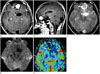
MRI revealed a large, well-demarcated, lobulated, heterogeneously enhanced, extra-axial mass with cystic areas in the left subfrontal area (Fig. 1A-C). Solid enhancement in the anterior portion and a non-enhanced cystic area in the posterior portion were noted. In addition, some peritumoral edema was seen. The solid enhanced portion showed diffusion restriction on diffusion MRI (Fig. 1D) and increased relative cerebral blood volume (rCBV) on perfusion MRI (Fig. 1E). However, the non-enhanced cystic portion did not show diffusion restriction on diffusion MRI or increased rCBV on perfusion MRI. MR spectroscopy showed increased choline/creatine peak and decreased N-acetylaspartate peak, though interpretation was limited due to serious baseline distortion.
Tumor excision was performed via bifrontal craniotomy. Intraoperatively, the tumor was located at the left olfactory groove, attached to the anterior part of the cribriform plate, and adherent to the dura. There were no procedure-related complications, and the tumor nearly disappeared on pre-discharge follow-up computed tomography. The patient was discharged home with partial improvement of symptoms.
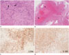
Histologic examination of the resected tissue revealed spindle-shaped cells with elongated nuclei and fibrillary cytoplasm (Antoni A pattern) (Fig. 2A), along with less cellular, loosely textured tumor areas (Antoni B). Extensive vascular hyalinization was seen (Fig. 2B), and immunohistochemical staining for CD56 (Fig. 2C), vimentin, and S100 (Fig. 2D) was positive. The pathologic diagnosis was confirmed as ancient schwannoma.
DISCUSSION
Intracranial schwannomas account for approximately 6% to 8% of all intracranial tumors (4). Schwannomas usually originate from the peripheral nerve sheath and occur less frequently in the central nervous system. Most intracranial schwannomas arise from the vestibulocochlear nerve, followed by the trigeminal nerve. Schwannomas with prominent degenerative change are referred to as ancient schwannomas, a very rare variant (3), which are characterized by diffuse hypocellular areas, relative loss of Antoni type A tissue, hyaline accumulation, calcification, cystic necrosis, hemorrhage, and fatty degeneration (5). This type of tumor may be misdiagnosed as malignancy due to its cellular degenerative changes. To date, there are only 36 cases of schwannoma in the anterior cranial fossa reported in the literature (1246789); moreover, there are no reports on the radiologic findings of ancient schwannoma.
Choi et al. (4) suggested that anterior cranial fossa schwannomas present different characteristics than lesions at other locations, such as earlier onset, male predominance, and a dominant solid tumor portion. Santhosh et al. (7) reported that schwannomas show multiple small hypointensities within the tumor on T2-weighted gradient-echo MRI sequence, which are presumed to be caused by microbleeds. In a literature review, Adachi et al. (6) reported that subfrontal schwannomas were solid and cystic and showed heterogeneous contrast enhancement. Luo et al. (10) suggested that extensive cystic degeneration is a characteristic finding of intracranial schwannoma not arising from the stems of the cranial nerves. Our patient showed similar findings, including an enhanced tumor with a cystic portion in the left subfrontal area, with microbleeds in the solid enhanced portion on T2-weighted gradient-echo MRI sequence. According to previous reports, extramural/arachnoid cysts may be caused by mechanical trapping of cerebrospinal fluid or by leakage of hemorrhagic material from the tumor, causing adhesion and secondary arachnoid cyst formation (9).
A previous report (1) suggested that the term "olfactory sch-wannoma" is incorrect because the olfactory bulb has no Sch-wann cell layer and is probably not the nerve of origin. Developmental and non-developmental hypotheses are suggested for the possible origin of olfactory schwannomas (8). According to developmental theories, mesenchymal pial cells are transferred to ectodermal Schwann cells, or neural crest cells migrate within the substance of the central nervous system (2). On the other hand, non-developmental theories suggest that schwannomas originate from the perivascular nerve plexus and meningeal branches of the trigeminal and anterior ethmoidal nerves, which innervate the anterior cranial fossa where Schwann cells are normally present (28).
Meningioma, esthesioneuroblastoma, metastasis, and others should be included in the differential diagnosis of extra-axial anterior cranial fossa neoplasm. Olfactory groove meningioma and schwannoma share similar radiologic features, including extra-axial location, calcification, enhancement pattern, and peritumoral edema. In our patient, the extra-axial mass was initially diagnosed radiologically as meningioma with peritumoral cyst based on its enhancement pattern and suspected dural tail sign on sagittal images. However, no definite bony erosion or sclerotic change of the cribriform plate was observed. Esthesioneuroblastoma was excluded from the differential diagnosis because it usually presents marked bony destruction. Metastasis was also excluded because of the unusual location, absence of extensive peritumoral edema, and the solitary mass.
Treatment options for schwannoma include surgical excision, observation, and radiotherapy, including Gamma knife surgery. However, complete tumor removal is the mainstay of treatment in order to prevent tumor recurrence. Moreover, patient prognosis after complete tumor resection is excellent.
In conclusion, schwannomas located at the olfactory groove are extremely rare and can be difficult to diagnose correctly before surgery. However, schwannoma should be considered in the differential diagnosis in cases of anterior cranial fossa neoplasm.
 XML Download