

 PDF
PDF ePub
ePub Citation
 Print
Print


INTRODUCTION
A retropharyngeal hematoma is an uncommon entity that may rapidly cause airway obstruction. The etiology of a retropharyngeal hematoma includes infections, cervical spine trauma, great vessel trauma, violent head movements, iatrogenic injury, parathyroid adenoma hemorrhage and a foreign body ingestion (1). The occurrence of a spontaneous retropharyngeal hematoma is rare and without any identifiable causes. In the majority of cases the diagnosis is delayed because of its rarity and absence of objective signs and diagnostic laboratory data. The author reports a case of spontaneous retropharyngeal hematoma in a 56-year-old man.
CASE REPORT
A 56-year-old man without a history of pre-existing neck disease presented to the emergency ward with sore throat, dysphonia and dyspnea. The symptoms were sudden in onset, severe in nature and rapidly progressive over two hours. The patient was a heavy alcoholics but had no prior cardiovascular, respiratory or gastrointestinal symptoms, no medical history or history of foreign body ingestion and also no past or current medication. The neck was non-tender and no significant limitation of neck movement was present. The subject was not febrile and presented a normal blood pressure. The hematologic evaluation showed a hematocrit of 47.9%, hemoglobin of 15.9 g/dL, white blood cell count with 13610/uL (neutrophil 73.7%, lymphocytes 15.7%, monocytes 8.2%), platelet count 251000/dL, erythrocyte sedimentation rate (ESR) 8 (0-10) mm/hr and C-reactive protein 25.3 (0-8) mg/L. Coagulation tests showed 96% prothrombin activity, a normal partial-thromboplastin time and a fibrinogen of 485 mg/dL. In the fiberoptic examination of the pharynx a significant anterior bulging of the posterior pharyngeal wall without ecchymosis was visible.
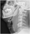
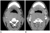
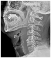
The plain neck lateral view showed a markedly increased thickness of the retropharyngeal pre-vertebral soft tissue (Fig. 1). On pre-contrast CT, the lesion which occupied the retropharyngeal space showed a slightly high-attenuation if compared with the adjacent muscle. In the region of interest in CT the numbers of lesions showed 75 Hounsfield unit (HU), which were higher than those of the adjacent muscle (55 HU). The post-contrast enhanced CT showed an expansile soft tissue attenuation at the retropharyngeal space with a slightly peripheral enhancement that ventrally displaced the posterior pharyngo-laryngeal wall with narrowing the airway from occiput to C7 level (Fig. 2). Three days later, a MR imaging was performed. On pre-contrast 91T1 weighted spin echo images [repetition time (TR)/echo time (TE), 433.3/10.0] a diffuse homogeneous hyperintensity was shown within the lesion. The lesion showed a low signal intensity on the fat suppressed T2 weighted images (TR/TE, 3500/47.9, short T1 inversion recovery). On the post-contrast T1 weighted fast spin echo images (TR/TE, 666.7/7.5) the lesion was not enhanced. Above mentioned image sequences disclosed typical signal characteristics of a subacute hematoma and represented intracellular methemoglobin converted from deoxyhemoglobin (Fig. 3) (2). The thickness of retropharyngeal prevertebral soft tissue was decreased in the 3 days follow-up check of the plain neck lateral view (Fig. 4).
The diagnosis of spontaneous retropharyngeal hematoma was suggested on the basis of clinico-radiologic findings. The patient remained stable with no progressive airway compromise. Conservative treatment was selected and a complete resolution of sore throat, dysphonia and dyspnea was reached. The patient was discharged within 5 days. The patient remained asymptomatic during the follow-ups 1 week and 1 month later.
DISCUSSION
Retropharyngeal hematoma is a rare entity with a fatal outcome potential owing to progressive internal blood loss and airway obstruction. The diagnosis can be difficult. In cases with no history of trauma, an early diagnosis in an outpatient department may be challenging because of non-specific-symptoms, such as neck pain or dysphagia, especially when a hematoma is limited to a retropharyngeal space. A patient may initially have only a sore throat without shortness of breath and may be misdiagnosed with viral pharyngitis (2). If a retropharyngeal mass is identified, the patient may be misdiagnosed with retropharyngeal abscess also.
The classical manifestations of cervicomediastinal hematomas are referred to as "Capps triad" and consist of tracheal and esophageal compression, anterior displacement of the trachea and subcutaneous bruising over the neck and anterior chest (2-6). The blood loss caused in a few cases a hypovolemic shock as a complication. However, in cases of moderate retropharyngeal hematoma, clinical signs are related to airway compression and include dysphagia and upper respiratory failure without a subcutaneous bruising (2).
The retropharyngeal hematoma is associated with a wide variety of etiologies. These include infection, cervical spine trauma, great vessel trauma, violent head movement, iatrogenic injury, parathyroid adenoma hematoma, foreign body ingestion and sudden pressure changes (due to vomiting, coughing and sneezing). Anticoagulation or hemorrhagic diathesis predisposes an individual to develop a retropharyngeal hematoma (1-3). A spontaneous retropharyngeal hematoma is defined by the absence of any clear etiology and any predisposing factor.
The retropharyngeal space is located immediately posterior to the naso-, oro- and hypopharynx, larynx and trachea. Its anterior border is formed by the buccopharyngeal fascia (surrounding the pharynx, trachea, esophagus and thyroid) and its posterior border is formed by the alar fascia. Laterally, it is bounded by the parapharyngeal space and the carotid sheaths (3-5, 7). The space consists of loose areolar tissue. A retropharyngeal hematoma is assumed to expand within this loose areolar tissue, which may delay symptoms for at least 2-3 hours and may possibly compromise the airway (3).
Plain radiography and CT and MRI are used to measure the prevertebral soft tissue thickness and to diagnose a retropharyngeal hematoma. Keats and Sistrom (8) reported that upper normal limits of normal range for thickness of prevertebral soft tissue in lateral cervical spine plain radiograph were 7 mm and 22 mm at C2 respective C6 levels. The upper normal limits on the multi detector CT were 6 mm at C2 and 18 mm at C6 and C7 levels (9). In the present case the prevertebral soft tissue thickness was increased to 38 mm at C3-C4 level on the neck CT. Using the multiplanar anatomic display and the tissue characterization CT and MR offer the exact localization of the lesion. The MR allows more specific diagnosis about blood products in different stages of evolution, because of their paramagnetic signal properties which change over the time depending on their dominant component (acute deoxyhemoglobins, subacute intra- or extracellular methemoglobins and chronic hemichromes) (2).
The differential diagnosis of retroperitoneal hematoma includes retropharyngeal infection, acute calcific prevertebral tendinitis and retropharyngeal effusion. Clinical and laboratory findings such as fever and dysphagia, leukocytosis and elevated ESR are important for the correct diagnosis. CT and MR images show a contrast enhancing retropharyngeal soft tissue or a ring enhancing abscess pocket formation. A patient with an acute calcified prevertebral tendinitis is less febrile and may have a normal white blood cell count. The CT of the such patient shows a calcific density in the site of the insertion of the longus colli muscle just anterior to the upper cervical spine. An effusion extends into the retropharyngeal space from C1 down to the level of C5 or C6. The abnormality begins in the prevertebral space rather than in the retropharyngeal space and shows less dense fluid collection without any contrast enhancement (10).
The treatment of retropharyngeal hematoma is basically to secure the airway and remove the hematoma. A close airway monitoring with the ability for an active intervention by intubation or a surgical maintenance of airway is essential in cases where an infection, foreign bodies or a continuous hematoma expansion are highly suggestive (2). However surgical intervention should be avoided unless a treatable etiological factor is found or an airway compromise occurs. Most cases of small or moderate hematomas can be resolved with a conservative management. The spontaneous retropharyngeal hematoma is usually less severe also (4).
The author presents a rare case of spontaneous retropharyngeal hematoma conservatively managed. A clinical diagnosis can be difficult for this potentially life-threatening condition. This case shows the possibility of an acute retropharyngeal swelling due to acute spontaneous hemorrhage. CT and MR imaging provide important informations for the correct diagnosis in this situation.

 XML Download