

 PDF
PDF ePub
ePub Citation
 Print
Print


Abstract
Cavernous hemangiomas of the mediastinum are rare tumors that usually occur in young patents. We present a case of cavernous hemangioma in a 12-year-old girl. This case was seen as nonenhancing cystic mass on CT, mimicking lymphangioma. US demonstrated a hyperechoic mass, while MRI revealed a T2 high SI with delayed homogenous enhancement. US and MRI were found to be useful tools for the diagnosis of hemangiomas, seen as a cystic mass on a chest CT scan.
Figures and Tables
Fig. 2
Chest CT images of a 12-year-old girl with mediastinal hemangioma.
A. Precontrast CT scan reveals low attenuating mediastinal mass (mean 40 HU) without phlebolith, surrounding great vessels and trachea.
B. Postcontrast CT scan shows little enhancement of the mass and right pleural effusion.
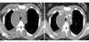
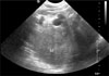
Fig. 4
Chest MRI images of a 12-year-old girl with mediastinal hemangioma.
A. Axial T1-weighted MRI shows heterogenous low to iso-intensity mass relatively hypointense to the skeletal muscle.
B. Axial T2-weighted MRI shows high-intensity mass with multiple structures of tubular signal voiding.
C. Axial dynamic enhanced T1-weighted MRI at 60 seconds reveals no enhancement of the mass.
D, E. Axial and coronal enhanced T1-weighted MRI after 60 minutes show diffuse, strong enhancement of the mass.

References
1. Cohen AJ, Sbaschnig RJ, Hochholzer L, Lough FC, Albus RA. Mediastinal hemangiomas. Ann Thorac Surg. 1987; 43:656–659.
2. Davis JM, Mark GJ, Greene R. Benign blood vascular tumors of the mediastinum: report of four cases and review of the literature. Radiology. 1978; 126:581–587.
3. McAdams HP, Rosado-de-Christenson ML, Moran CA. Mediastinal hemangioma: radiographic and CT features in 14 patients. Radiology. 1994; 193:399–402.
4. Semisa M, Fiore D, Ravasini R, Macchi C. Computed tomography of mediastinal cavernous hemangioma: a case report. Rays. 1986; 11:31–33.
5. Bree RL, Schwab RE, Glazer GM, Fink-Bennett D. The varied appearances of hepatic cavernous hemangiomas with sonography, computed tomography, magnetic resonance imaging and scintigraphy. Radiographics. 1987; 7:1153–1175.
6. Agarwal PP, Seely JM, Matzinger FR. Case 130: mediastinal hemangioma. Radiology. 2008; 246:634–637.
7. Fujimoto K, Muller NL. Anterior mediastinal masses. In : Muller NL, Silva CI, editors. Imaging of the chest, volume II. Philadelphia: Saunders;2008. p. 1512.
8. Ishii K, Maeda K, Hashihira M, Miyamoto Y, Kanegawa K, Kusumoto M, et al. MRI of mediastinal cavernous hemangioma. Pediatr Radiol. 1990; 20:556–557.
9. Abe K, Akata S, Ohkubo Y, Park J, Kakizaki D, Simatani H, et al. Venous hemangioma of the mediastinum. Eur Radiol. 2001; 11:73–75.
10. Nchimi A, Ghaye B, Szapiro D, Thiry A, Dondelinger RF. A complex anterior mediastinal mass: demonstration of pericardial haemangioma by dynamic MRI (2003:10b). Eur Radiol. 2004; 14:160–163.
 XML Download