Abstract
Background:
Triphasic waves are one of the electroencephalographic patterns that can be usually seen in metabolic encephalopathy. The aim of this study is to compare the clinical and electrophysiologic profiles between patients with and without triphasic waves in metabolic encephalopathy, and reassess the significance of triphasic waves in metabolic encephalopathy.
Methods:
We recruited 127 patients with metabolic encephalopathy, who were admitted to our hospital. We divided these admitted patients into two groups; those with and without triphasic waves. We analyzed the difference of duration of hospitalization, mortality rate during admission, Glasgow Coma Scale, severity of electroencephalographic alteration, and presence of acute symptomatic seizures between these two groups.
Results:
Of the 127 patients with metabolic encephalopathy, we excluded 67 patients who did not have EEG, and 60 patients finally met the inclusion criteria for this study. Patients with triphasic waves had more severe electroencephalographic alterations, lower Glasgow Coma Scale, and more acute symptomatic seizures than those without triphasic waves. After adjusting the clinical variables, Glasgow Coma Scale and acute symptomatic seizures were only significantly different between patients with and without triphasic waves.
REFERENCES
2.Kaplan PW. The EEG in metabolic encephalopathy and coma. J Clinical Neurophysiol. 2004. 21:307–318.
3.Bickford RG., Butt HR. Hepatic coma: the electroencephalographic pattern. J Clin invest. 1955. 34:790–799.

5.Sundaram MB., Blume WT. Triphasic waves: clinical correlates and morphology. Can J Neurol sci. 1987. 14:136–140.

6.Young GB., Bolton CF., Austin TW., Archibald YM., Gonder J., Wells GA. The encephalopathy associated with septic illness.
7.Aguglia U., Gambardella A., Oliveri RL., Lavano A., Quattrone A. Nonmetabolic causes of triphasic waves: a reappraisal. Clin Electroencephalogr. 1990. 21:120–125.

8.Konno S., Sugimoto H., Nemoto H., Kitazono H., Murata M., Toda T, et al. Triphasic waves in a patient with tuberculous meningitis. J Neurol Sci. 2010. 291:114–117.

9.Foley JM., Watson CW., Adams RD. Significance of the electroencephalographic changes in hepatic coma. Trans Am Neurol Assoc. 1950. 51:161–165.
10.Bahamon-Dussan JE., Celesia GG., Grigg-Damberger MM. Prognostic significance of EEG triphasic waves in patients with altered state of consciousness. J Clin Neurophysiol 1989,6;. 313–319.
11.Beghi E., Carpio A., Forsgren L., Hesdorffer DC., Malmgren K., Sander JW, et al. Recommendation for a definition of acute symptomatic seizure. Epilepsia. 2010. 51:671–675.

12.Kaplan PW., Schlattman DK. Comparison of triphasic waves and epileptic discharges in one patient with genetic epilepsy. J Clin Neurophysiol. 2012. 29:458–461.

13.Boulanger JM., Deacon C., Lecuyer D., Gosselin S., Reiher J. Triphasic waves versus nonconvulsive status epilepticus: EEG distinction. Can J Neurol sci. 2006. 33:175–180.

14.Chong DJ., Hirsch LJ. Which EEG patterns warrant treatment in the critically ill? reviewing the evidence for treatment of periodic epileptiform discharges and related patterns. J Clin Neurophysiol. 2005. 22:79–91.

15.Nowack WJ., King JA. Triphasic waves and spike wave stupor. Clin Electroencephalogr. 1992. 23:100–104.

16.Andraus ME., Andraus CF., Alves-Leon SV. Periodic EEG patterns: importance of their recognition and clinical significance. Arq Neuropsiquiatr. 2012. 70:145–151.

17.Kuroiwa Y., Celesia GG. Clinical significance of periodic EEG patterns. Arch Neurol. 1980. 37:15–20.

18.Beghi E., Tognoni G. Prognosis of epilepsy in newly referred patients: a multicenter prospective study of the effects of mon-otherapy on the long-term course of epilepsy. Collaborative Group for the Study of Epilepsy. Epilepsia. 1992. 33:45–51.
19.Sillanpaa M., Jalava M., Kaleva O., Shinnar S. Long-term prognosis of seizures with onset in childhood. N Engl J Med. 1998. 338:1715–1722.

20.Lehembre R., Gosseries O., Lugo Z., Jedidi Z., Chatelle C., Sadzot B, et al. Electrophysiological investigations of brain function in coma, vegetative and minimally conscious patients. Arch Ital Biol. 2012. 150:122–139.
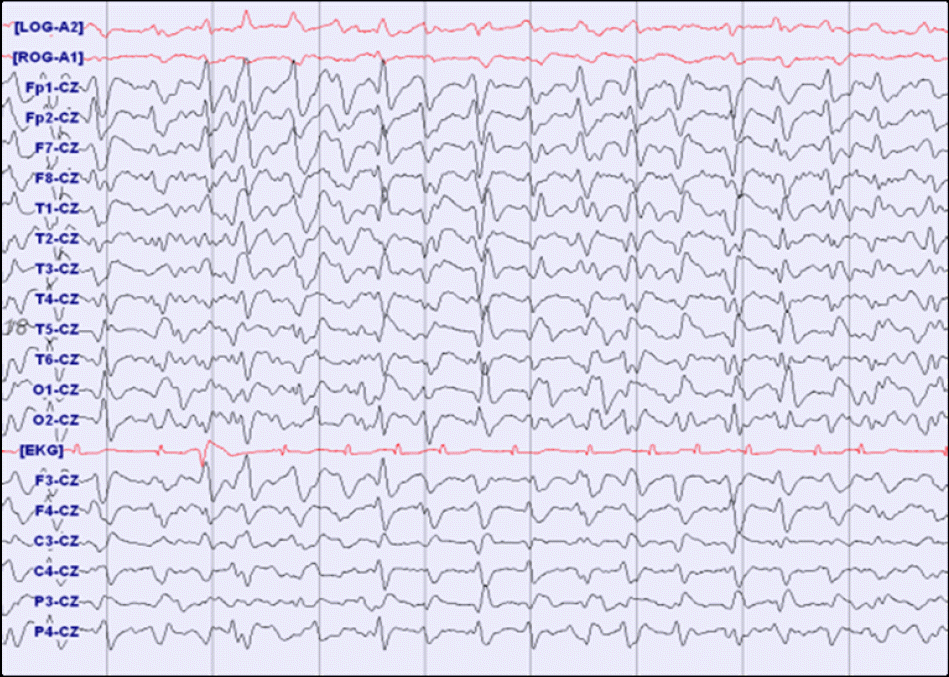
Figure 1.
An electroencephalography of the 85-year-old woman with acute renal failure shows bilateral synchronous, frontally dominant runs with waveforms of phase: negative, positive, negative, and fronto-occipital lag.

Table 1.
A comparison of the clinical and electrophysiologic profiles between patients with and without triphasic waves in metabolic encephalopathy


 PDF
PDF ePub
ePub Citation
 Print
Print


 XML Download