

 PDF
PDF ePub
ePub Citation
 Print
Print


Abstract
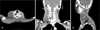
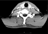
Pseudomeningoceles are formed by extravasation of cerebrospinal fluid through a dural defect into soft tissue. Most pseudomeningoceles are iatrogenic and occur in the posterior lumbar region following surgery. But, post-traumatic pseudomeningocele rarely occurs in the head and neck. We present a case of post-traumatic an unusual cervical pseudomenigocele and review the related literatures.
Figures and Tables
References
1. Andrew SA, Sidhu KS. Cervical-peritoneal shunt placement for postoperative cervical pseudomeningocele. J Spinal Disord Tech. 2005; 18:290–292.
2. Hawk MW, Kim KD. Review of spinal pseudomeningoceles and cerebrospinal fluid fistulas. Neurosurg Focus. 2000; 9:e5.

3. Horn EM, Bristol RE, Feiz-Erfan I, Beres EJ, Bambakidis NC, Theodore N. Spinal cord compression from traumatic anterior cervical pseudomeningoceles. Report of three cases. J Neurosurg Spine. 2006; 5:254–258.
4. Hosono N, Yonenobu K, Ono K. Postoperative cervical pseudomeningocele with herniation of the spinal cord. Spine (Phila Pa 1976). 1995; 20:2147–2150.
5. Jeong JH, Ahn SK, Jeon SY, Park JJ, Kim JP, Park IS. Post-traumatic pseudomeningocele presenting as a cyst of external auditory canal: report of a case. Auris Nasus Larynx. 2006; 33:321–324.
6. Kitchen N, Bradford R, Platts A. Occult spinal pseudomeningocele following a trivial injury successfully treated with a lumboperitoneal shunt: a case report. Surg Neurol. 1992; 38:46–49.
7. Lau KK, Stebnyckyj M, McKenzie A. Post-laminectomy pseudomeningocele: an unusual cause of bone erosion. Australas Radiol. 1992; 36:262–264.
8. Maycock NF, van Essen J, Pfitzner J. Post-laminectomy cerebrospinal fluid fistula treated with epidural blood patch. Spine (Phila Pa 1976). 1994; 19:2223–2225.
9. McCormack BM, Taylor SL, Heath S, Scanlon J. Pseudomeningocele/CSF fistula in a patient with lumbar spinal implants treated with epidural blood patch and a brief course of closed subarachnoid drainage. A case report. Spine (Phila Pa 1976). 1996; 21:2273–2276.
10. Naso WB, Cure J, Cuddy BG. Retropharyngeal pseudomeningocele after atlanto-occipital dislocation: report of two cases. Neurosurgery. 1997; 40:1288–1290.
11. Rinaldi I, Hodges TO. Iatrogenic lumbar meningocoele: report of three cases. J Neurol Neurosurg Psychiatry. 1970; 33:484–492.
12. Ryall RG, Peacock MK, Simpson DA. Usefulness of beta 2-transferrin assay in the detection of cerebrospinal fluid leaks following head injury. J Neurosurg. 1992; 77:737–739.
13. Shapiro SA, Scully T. Closed continuous drainage of cerebrospinal fluid via a lumbar subarachnoid catheter for treatment or prevention of cranial/spinal cerebrospinal fluid fistula. Neurosurgery. 1992; 30:241–245.
14. Tate S, Rak RA, Bailey JS. Unusual presentation of a cervical pseudomeningocele: a case report and review of the literature. J Oral Maxillofac Surg. 2005; 63:556–559.
15. Taveras JM, Ransohoff J. Leptomeningeal cysts of the brain following trauma with erosion of the skull; a study of seven cases treated by surgery. J Neurosurg. 1953; 10:233–241.
 XML Download