

 PDF
PDF ePub
ePub Citation
 Print
Print


INTRODUCTION
The osteotome sinus floor elevation (OSFE) introduced by Summers [1], is generally used in moderately resorbed posterior maxilla to graft the maxillary sinus in combination with immediate implant placement [2]. This technique requires striking the bone with a surgical mallet until the desired depth is reached.
During the installation of maxillary dental implants using the OSFE, the surgical trauma induced by percussion with the surgical mallet, along with hyperextension of the neck during the operation, can displace otoliths and result in the appearance of benign paroxysmal positional vertigo (BPPV) [3]. BPPV is a common vestibular end organ disorder characterized by short, often recurrent episodes of vertigo that are triggered by certain head movements in the plane of the posterior semicircular canals [4].
Although the incidence of OSFE-induced BPPV was less than 3% (4 in 146 patients) [5] and commonly resolves itself within a month without treatment [2], the symptoms involved are very unpleasant [4]. The symptoms are sufficiently severe to significantly hinder patients from carrying out normal daily activities if not identified correctly and managed properly.
A case of BPPV in a young man caused by surgical trauma during implant placement performed with the use of osteotomes is reported in this article.
CASE DESCRIPTION
This case report was exempted from the approval of the institutional review board. A healthy 27-year-old Korean man visited the Department of Periodontics in the Gangneung-Wonju National University Dental Hospital for implant placement in the edentulous region of the maxillary right first molar. The patient was in good physical health with no history of sinus disease or vertigo. He was not taking medications and denied any allergies. The maxillary right first molar had been extracted due to severe dental caries approximately 10 years prior to presentation.
Panoramic radiographs were taken for evaluation of the maxillary sinus and general dental state. Since radiographic evaluation revealed that the bone height between the alveolar ridge crest and sinus floor was 8 mm, the bone-adding OSFE procedure with simultaneous implant installation was planned. The patient was prepared in a routine aseptic manner.
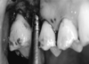
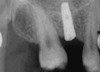
Surgery was performed under local anesthesia (approximately 6 mL of 2% lidocaine hydrochloride with 1:100,000 epinephrine, Lignospan standard, Septodont, Cedex, France). The osteotomy was initiated with a guide drill to penetrate the cortical bone, followed by a 2 mm twist drill drilling to 1 mm short of the sinus floor. The cortical plate of the sinus floor was fractured for the bone-adding OSFE procedure using a surgical mallet and increasingly wider osteotomes. Because the cortical bone plate of the sinus floor was very hard, heavy malleting on osteotomes was done to elevate the sinus floor to the desired depth. Granular anorganic bovine bone measuring 0.25 to 1 mm (Bio-Oss®, Geistlich AG, Wollhusen, Switzerland) was intermittently added to the implant osteotomy site during the sinus elevation procedure (Fig. 1). A 4 × 10 mm implant (Shinhung Implant M, Tapered type, Shinhung, Seoul, Korea) was then placed in the osteotomy site (Fig. 2). The surgery lasted about 40 minutes. Upon sitting up after surgery, the patient experienced intense vertigo with nausea, especially when he changed the position of his head. He sat in the dental chair and rested for 30 minutes, but he was still affected by vertigo.
The next day, the patient was referred to an otorhinolaryngologist for treatment of the postoperative vertigo. After a positive Dix-Hallpike test, posterior canal BPPV at the right inner ear was diagnosed. During the Dix-Hallpike test, a nystagmus response and vertigo was provoked on the cephalic hyperextension with right lateralization. The Epley maneuver [6] was performed once on the patient's right side, and anti-vertigo drugs (dimenhydrinate 50 mg, Bonaling-A®, Il Yang Pharm., Seoul, Korea) were prescribed to help him maintain a normal life. Nevertheless, the patient stayed vertiginous for 1 week. On follow-up dental visits at 7 and 8 days, an implant surgeon performed additional Epley maneuvers. After the Epley maneuvers by the dentist, the patient became asymptomatic; he remained asymptomatic 1 year later.
DISCUSSION
Recently, the incidence of posterior maxilla implant placement is increasing in the elderly population. With increasing age, the resorptive process predominates over new bone formation, and the loss of teeth leads to increased osteoclastic activity, resulting in bone height deficiency in the alveolar process of the maxilla [5]. When there is less than 10 mm of bone remaining between the crest of the maxilla and the floor of the sinus, bone augmentation must be considered [1]. Tatum [7] introduced the crestal approach and lateral approach sinus augmentation procedures in the 1970s [1]. In the crestal approach, a special set of osteotomes (Summers Osteotome Kit) and a surgical mallet were used to raise the sinus floor [1].
Preparation of the implant beds with osteotomes and a surgical mallet transmits percussive forces capable of detaching heavy, inorganic particles (otoliths) from the otoconia layer of the utricular macula [2,4,8]. Moreover, during surgical positioning of the patient face up and his head in hyperextension favor the displacement of otoliths into the posterior semicircular canal [2-5] (Canalithiasis ; i.e. that which is free-floating from the macula and gravitates into the endolymph of the semicircular canal). Then the otoliths float around in the gravity-sensitive endolymph system, causing vertigo [8]. These disturbances are almost in the plane of the posterior semicircular canal, although in some cases the other semicircular canal is affected [3].
The diagnosis of BPPV is established by inducing a rapid change from the sitting position to the left or right head-hanging position - the Dix-Hallpike test [2,9,10]. Patients with BPPV experience vertigo when moved rapidly into a supine position with the head turned so that the affected ear is 30 to 45 degrees below the horizontal plane [2,10,11]. The vertigo occurs with a latency of 1 to 40 seconds after the patient has been placed in the provoked position (usually after 1 to 5 seconds) [2,10,11]. The patient also develops a characteristic nystagmus, which is torsional with the eyes directed toward the affected side, becoming more vertical ("up beating") when the eyes are directed away from the affected side [2,10,11]. The vertigo and nystagmus then disappear in approximately 30 to 60 seconds [2,10,11]. It should be noted that patients who develop vertigo with this maneuver but do not develop the nystagmus do not have BPPV [10].
Although BPPV is a "self-limiting" disorder and commonly resolves itself within a month [10], the symptoms involved are very unpleasant for the patient. Therefore, implant surgeons must resolve this condition quickly with appropriate treatment.
The symptoms of vertigo due to many different underlying etiologies are commonly treated with medications [6]. Clinicians may prescribe pharmacological management to either reduce the spinning sensation of vertigo and/or to reduce the accompanying motion sickness symptoms. The most commonly used are benzodiazepines and antihistamines. Benzodiazepines, such as diazepam, and clonazepam, have anxiolytic, sedative, muscle relaxant, and anticonvulsant properties derived from potentiating the inhibitory effect of the gamma-amino butyric acid system. Antihistamines, on the other hand, appear to have a suppressive effect on the central emetic center which relieves the nausea and vomiting associated with motion sickness. Common examples of antihistamines used to treat symptoms of vertigo and/or associated motion sickness include meclizine and diphenhydramine. Motion sickness medications are sometimes helpful in controlling the nausea, but are generally not very beneficial [6,8].
Physical maneuvers have proven to be very effective in treating BPPV. The canalith repositioning procedure, known as the "Epley maneuver," which is used in posterior semicircular canal BPPV cases, is effective in about 80% of patients [6,8,11,12]. Through a series of head position changes, the Epley maneuver moves the canaliths from the posterior semicircular canal to the vestibule, thereby relieving the stimulus from the semicircular canal that had been producing the vertigo in BPPV [6]. Following repositioning of the canaliths, the subject of this report has had no recurrence of his symptoms.
This report shows that the clinician should know about and expect BPPV after the osteotome technique. To prevent this complication, gentle malleting and a careful approach should be taken during the osteotome technique. The lateral approach and the use of other surgical kits for a crestal approach instead of mallet percussion should prevent the trauma to the craniofacial area as much as possible. In suspected cases of BPPV, the patient should be informed about the possibility of postoperative vestibular symptoms because these symptoms may be very unpleasant and may cause considerable stress when one is unaware of this problem. As the symptoms may be very incapacitating, immediate referral to an otorhinolaryngologist is strongly recommended. Early diagnosis of BPPV and immediate application of the Epley maneuver may help reduce the patient's discomfort.
 XML Download