

 PDF
PDF ePub
ePub Citation
 Print
Print


To the editor:
Chronic lymphocytic leukemia (CLL) is a malignant proliferation and accumulation of abnormal mature lymphocytes. Though extranodal involvement is common, vulovaginal involvement by CLL is extremely uncommon, especially as the primary presentation. This case is presented for its rarity and to highlight the importance of exact pathologic diagnosis in the management of these patients.
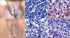
A 55-year-old female was evaluated for swelling and ulcerative lesion of the vulva for which she had consulted two separate gynecologists and had been treated with topical antifungals and antibiotics without any improvement. She presented to us with a persistent vulval lesion of 8 months duration. By this time she had also developed bilateral neck nodes enlargement of 2 months duration. On examination she had generalized lymphadenopathy and hepatomegaly. Detailed gynecological examination was performed and an ulcerative lesion with rolled out edges was found in the inner surfaces of both labia majorae (Fig. 1A). Speculum examination revealed an inflamed vaginal mucosa with prominent mucosal folds. The cervix was unhealthy with mucosal irregularity. Investigations revealed a haemoglobin of 98 gm/L, total white cells of 110×109/L with differential count showing lymphocyte of 88%, and platelet count of 359×109/L. Her bone marrow examination and flow cytometry was suggestive of CLL. The patients' labial biopsy showed sheets of mature appearing lymphocytes which was CD 5, CD 20, CD 23, LCA and CD 79α, CD 43 positive (Fig. 1B, C, E) and the endocervical smear also showed many mature appearing lymphoid cells suggestive of CLL involvement (Fig. 1F). The patient has been treated with 2 cycles of bendamustine and is responding well to treatment with significant regression of the vulval swelling (Fig. 1D).
CLL is commonly diagnosed incidentally in asymptomatic patients when a routine physical examination reveals non-tender lymphadenopathy, or routine blood investigation shows unexplained absolute lymphocytosis. CLL can also present as involvement of extralymphoid organ, and skin being most common, accounting for 8% of cases [1]. Female genital tract involvement can occur in Non-Hodgkin's lymphomas (NHLs) and the most common is diffuse large B cell lymphoma [2]. Very few cases of vulvovaginal involvement of CLL have been reported in the English literature and this is only the second case of CLL with involvement of vulva as well as cervix [2-4].
In addition to carcinoma of the vagina/vulva, many unusual cancers have been reported to involve these areas. The differential diagnosis vulvovaginal lesions which needs to be kept in mind by practicing gynecologists and physicians include vaginal NHL and other hematopoietic lesions (granulocytic sarcoma, Langerhan's cell histiocytosis), malignant mixed Mullerian tumor, epithelioid leiomyosarcoma, endometrial stromal tumors including endometrial stromal sarcoma, melanoma, and extraosseous Ewing's sarcoma/primitive neuroectodermal tumor [5].
Our patient initially presented with swelling and ulceration in vulva, the diagnosis of which was missed due to rarity of the presentation. She was diagnosed as having CLL eight months later, only after she developed generalized lymphadenopathy. It is beneficial to identify patients with CLL in its early stages so that they may be monitored for disease progression, and therapy can be initiated as soon as indicated. In our case, early pick of the CLL would have been possible if the gynecologist performed a biopsy when the vulval lesion had failed to respond to antifungal and antibiotic treatment. We present this case for its rarity and to highlight the importance of early biopsy in gynecological lesions not responding to conventional therapy.
 XML Download