

 PDF
PDF ePub
ePub Citation
 Print
Print


INTRODUCTION
Recipients of solid organ transplants have an increased risk of developing certain types of malignancies as compared with that of the general population [1]. Liver transplantation has been increased over the last two decades in Korea, and its good outcome with long term survival has prompted an increase of post-transplant complications [2]. Among the neoplasms that can develop after liver transplantation, post transplant lymphoproliferative disorder (PTLD), lymphoma, malignant melanoma, cutaneous basal-cell and squamous cell carcinoma (SCC), and SCC in situ of the vulva have been reported [3].
Here we report on the first pediatric Korean case of widely spread-vulvar SCC in situ that developed 14 years after living-related donor liver transplantation. The present case is unique in that the vulvar SCC in situ was associated with infection by uncommon human papillomavirus (HPV) subtypes as well as high risk subtypes.
CASE REPORT
A 16-year-old girl presented with multiple verruocoid vulvar masses on both the labia majora and minora. The lesions were incidentally detected 16 months previously. She did not complain of local symptoms. No gross abnormalities were found in the vagina. She denied any sexual intercourse history, but her hymen was torn. At the age of 18 months, she underwent living-related donor liver transplantation from her mother due to neonatal hepatitis and subsequent cirrhosis. After that, she had been taking immunosuppressive agents, mainly tacrolimus (FK506, 2 mg daily). She experienced two bouts of acute cellular rejection in superimposed chronic advanced rejection. Steroid pulse therapy and an increased dosage of tacrolimus were administered each time. Two years earlier, a high fasting glucose level (160 mg/dL) was noticed on the routine follow-up. Diabetes mellitus was diagnosed, and Novorapid (Aspart, Pen, 300 IU/3 mL per day) therapy was taken. She had been followed up every two months during the 14 years of the postoperative follow-up period. The laboratory findings, including the peripheral blood cell counts and except for the hyperglycemia, were within the normal levels.
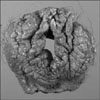
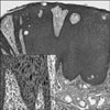
Wide excision of the bilateral labia majora and minora with skin grafts was done. The excised specimen showed that the largest cauliflower-shaped polypoid lesion measured 3.5×2.0×0.3 cm (Fig. 1). The vulvar lesions showed various degrees of vulvar intraepithelial neoplasm II and III. Some small uniform basaloid cells showed hyperchromatic and coarse nuclear chromatin, and the warty larger cells showed greater pleomorphism with koilocytotic changes near the surface (Fig. 2). Nonkeratinizing, basaloid type of SCC in situ was also found. A cervicovaginal smear showed multiple koilocytotic dysplastic cells, and this was diagnosed as low grade squamous intraepithelial lesion with HPV infection and mild dysplasia.
We also analyzed the HPV by a PCR-based DNA microarray system provided by a HPV-DNA chip test (MyHPVChip; Biomedlab Co. Ltd., Seoul, Korea) and using paraffin blocks. The chip contains 32 type-specific probes that consist of 18 high-risk HPV groups (16, 18, 26, 31, 33, 35, 39, 45, 51, 52, 56, 58, 59, 66, 68, 69, 70, 73) and 14 low-risk HPV groups (6, 11, 32, 34, 40, 42, 43, 44, 53, 54, 55, 57, 61, 62). HPV types 69 and 73 and HPV types 66, 70, 73, and 43 were identified in the vulvar mass and the cervicovaginal smear, respectively. She had an uneventful postoperative recovery. There has been no recurrence during the 3 months of follow up and her blood glucose level is under control.
DISCUSSION
Liver transplantation is the standard of care for children with end-stage, irreversible liver disease. As with adults, the early major complications include rejection, opportunistic infection and PTLD, and especially in children [4]. Post-transplant diabetes mellitus, as shown in the present patient, is one of the frequent complications after liver transplantation, and it develops in 10% to 30% of all these patients [3]. The mechanism of pediatric post-transplant diabetes is likely to be a result of both insulin deficiency and the insulin resistance of immunosuppressive agents, including steroids and tacrolimus [5]. After a long follow-up duration, there is an increased incidence of certain malignancies developing; the relative risk for malignancies is two to three times higher than that in the general population [1,2]. The most common malignancies in pediatric transplant recipients are lymphomas and skin and lip carcinomas, whereas skin and lip carcinomas are the most common in adults [6]. Vulvar SCC usually appears in postmenopausal women in the 7th and 8th decades of life. Despite the young age, the multicentric squamous intraepithelial lesion (vulva and uterine cervix) was detected as well as HPV-infection in the present case. Post-transplant recipients demonstrated a 25- to 40-fold increased risk to develop vulvar carcinomas [7]. About 40% of vulvar SCCs are associated with HPV-infection. In one study, 100% of the vulvar lesions in post-transplant recipients were HPV-positive compared to 21-57% in immunocompetent individuals [8]. Immunosuppression per se is a known risk factor for HPV oncogenic virus-disease, and immunosuppressive agents and HPV play synergistic roles in carcinogenesis [9]. HPV is widely accepted as a causative agent of anogenital cancer. To date, more than 118 HPV genotypes have been identified and sequenced, and about 40 can infect the genital tract and 12 are classified as oncogenes [10,11]. By their oncogenic potential, HPV types were classified as high risk (16, 18, 31, 33, 35, 39, 45, 51, 52, 56, 58, 59, 68, 73, and 82), low risk (6, 11, 40, 42, 43, 44, 54, 61, 70, 72, 81, and CP6108) and probable high risk (26, 53, and 66). HPV type 16 has previously shown to be the most common type in all countries including Korea [12]. However, HPV type 69, which is one of the subtypes detected in our patient's vulvar carcinoma, is previously unknown in genital SCC. A recent world-wide assessment of HPV genotypes has shown that the previously unknown HPV types, which were identified as type 26, 30, 61, 67, 69, 82, and 91, comprise 1% of all the studied invasive cervical carcinomas [13]. The biological behaviors of the rare and uncommon HPV types have only been investigated in a few studies, and mostly included lesions that were cervical intraepithelial neoplasms and a few cases of invasive cervical cancer [9]. Little is known about the exact mechanism of HPV-associated vulvar carcinogenesis of these rare HPV types because of the insufficient epidemiological evidence. HPV vaccination is prophylactic, and it is assumed that maximum effectiveness will be achieved by administration before exposure to HPV or viral reactivation because HPV vaccination may offer a boosting effect of previously acquired natural immunity [14]. Two recent vaccines, the recombinant quadrivalent HPV vaccine (Merck, Gardasil®) and the bivalent HPV recombinant vaccine (GalxoSmithKline, Cervarix®), can mainly prevent the HPV targeting types 16, 18, 6, 11, 31, and 9 for Gardasil and types 16, 18, 45, and 31 for Cervarix. However, these vaccines are not effective in protecting against other high-risk HPV types, including the above mentioned uncommon types, which were also found in the present case.
In conclusion, the present case is the first reported Korean case of vulvar SCC that developed in an adolescent who was taking immunosuppressive drugs after liver transplantation. Specialized vaccination programs may be required in post-transplant recipients because uncommonly found HPV subtypes may be ascribed to the carcinogenesis of vulvar SCC, especially in immunocompromised patients. This case is the first to show that uncommon HPV subtypes are related to pediatric post-transplant vulvar SCC. Albeit the incidence of pediatric vulvar SCC is rare, physicians should carefully perform more regular periodic genital examinations in children and adolescent transplant recipients because of their long-term follow-up and improved survival.
 XML Download