

 PDF
PDF ePub
ePub Citation
 Print
Print


INTRODUCTION
Malignant transformation of mature cystic teratoma (MCT) of the ovary is very rare (1.8%).1 The most common transformation within a mature cystic teratoma is squamous cell carcinoma (75-83%), and adenocarcinoma arising from mature cystic teratoma is extremely rare, accounting for just 6.8%. The diagnosis of mature cystic teratoma is relatively easy because of specific radiologic findings and such a benign mass is often removed by laparoscopic operation. Although some previous studies reported that patients who developed malignant transformation were usually postmenopausal and symptomatic with abdominal pains,2 pre-operative diagnosis of malignant transformation is difficult. Therefore, patients should be informed that a prompt second staging operation should be performed if the definitive pathology reveals an unexpected malignancy after laparoscopic cystectomy.3 In this paper, we present a case of a patient with thyroid papillary carcinoma of follicular variant arising from a mature cystic teratoma removed by laparoscopic salpingo-oophorectomy, followed by staging laparotomy.
CASE REPORT
A 35-year-old multiparous (para 2-0-2-2) woman presented with intermittent low abdominal discomfort lasting several months prior to her visit. She complained of mild constipation and indigestion.
In the initial pelvic examination, an approximately double fist-sized soft cystic mass was detected in the pelvic cavity. Transvaginal ultrasonography revealed a 16.7×11.5 cm sized multiseptated cystic mass on left ovary. These findings suggested the presence of a cystadenoma or dermoid cyst. Whole blood count, urine analysis, liver function tests were unremarkable. The tumor marker levels were as follows-carcinoembryonic antigen (CEA), 1.0 ng/ml; alpha-fetoprotein (AFP), 2.5 ng/ml; CA19-9, 11.5 IU/ml; CA125, 31.3 U/ml. Pelviscopic operation was planned with the provisional diagnosis of a benign ovarian cyst.
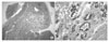
Exploratory pelviscopy revealed a 13×11 cm, whitish, well capsulated, mobile left ovarian cyst. No ascites was detected. After aspiration of the internal cyst fluid which was composed of yellowish serous fluid, left salpingo-oophorectomy was done. The tumor mass consisted of yellowish gray fibromembranous tissues and was removed in multiple fragments. The final pathological diagnosis is papillary carcinoma, follicular variant arising from a mature cystic teratoma of the left ovary (Fig. 1). Thyroid transcription factor-1 and thyroglobulin was expressed in the tumor cells. Two months later, staging operation including total abdominal hysterectomy and pelvic lymph node dissection was done, and which showed no residual tumors grossly. There was also no malignant findings in the final pathologic report. The patient had no evidence of disease 6 months after the operation.
DISCUSSION
Benign or mature cystic teratomas, commonly called dermoid cysts, are composed of mature tissues, which can contain elements of all three germ cell layers. They are recognized as one of the most common tumors in women during the reproductive age.
Malignant transformation of a benign cystic teratoma is a rare event. Although preoperative diagnosis of epithelial ovarian malignancy is not so difficult, distinguishing this malignant transformation from benign cystic teratoma preoperatively is nearly impossible. Many tumor markers such as carcinoembryonic antigen (CEA), CA 125, CA 19-9, human chorionic gonadotrophin (hCG), alpha fetoprotein (AFP), and lactate dehydrogenase (LDH) may be useful in diagnosing ovarian malignancy, but they are nonspecific and cannot exclude ovarian malignancy entirely.4 Mori et al. studied retrospectively the ability to diagnose malignant transformation of teratomas preoperatively.5 They concluded that among age, tumor size, serum CEA levels, and serum SCC levels, the combined two criteria of patient's age (under 40 years old or younger) and serum SCC level (under 2.5 ng/ml) is a suitable marker for differential diagnosis between benign mature cystic teratomas and malignant transformation. Kawai et al.6 studied seven tumor markers in ovarian tumors and reported that CA 19-9 showed a high positive rate in teratomatous tumors which were immature teratoma, mature cystic teratoma with malignant transformation, and mature cystic teratoma. Kikkawa et al.7 reported that age and tumor size are important factors in making a differential diagnosis, and recommended that serum SCC and CEA levels should be measured in patients older than 45 years who have a mature cystic teratoma-like ovarian tumor larger than 99 mm in the greatest diameter. But these reports pertain to squamous cell carcinomas arising in mature cystic teratomas, which is the most common type of malignant transformation. There were few studies for adenocarcinoma arising in mature cystic teratoma.
Adenocarcinoma arising from benign mature cystic teratomas is extremely rare and in 1957, Peterson et al. reported only 48 cases in the literature. Because of the rarity of these cancers, few studies have been able to make a pre-operative diagnosis and which might be overlooked as a benign teratoma with normal SCC and CEA level. As mentioned above, squamous cell carcinomas arising in a mature cystic teratoma might be suspected because of elevated SCC levels and large tumor size, but in adenocarcinomas arising from a mature cystic teratoma, there were no studies describing tumor markers or size. Among 9 cases of adenocarcinoma in which tumor markers were measured, including this case, only 2 cases showed elevated serum CEA level, 3 cases with elevated CA 19-9 level, and 2 cases with elevated CA125 level.8-12 The size of tumor varied from a smallest tumor size of 4 mm in diameter to 36 cm.8 These results reveal that specific tumor markers and tumor sized are ineffective in contributing to the preoperative diagnosis of adenocarcinomas arising from mature cystic teratoma. The tissue origin of these reports was various such as sweat glands, salivary glands, mammary glands, respiratory ciliated epithelium, gastrointestinal and thyroid glands.10,11,13,14 In this study, we could find the tissue origin of thyroid transcription factor-1 and thyroglobulin by immunohistochemistry.
In conclusion, we present a premenopausal woman with thyroid papillary carcinoma of follicular variant arising from mature cystic teratoma with normal tumor makers treated by laparoscopic salpingo-oophorectomy followed by staging laparotomy.
 XML Download