

 PDF
PDF ePub
ePub Citation
 Print
Print


Introduction
Endoscopic submucosal dissection (ESD) is accepted as the primary treatment for early gastric cancer (EGC) lesions that meet the absolute indications for resection and can be considered as an investigational treatment for lesions that meet the expanded indications and have a negligible risk of lymph node metastasis.1,2 After ESD for EGC lesions that meet the final pathological curability criteria for ESD indications, long-term outcomes are favorable and comparable between both criteria for absolute and expanded indications. The 5-year overall survival (OS) rate is 92% to 97% for the patients with absolute indications and 93% to 97% for the patients with expanded indication.3,4,5 Furthermore, the OS after endoscopic resection (ER) was comparable with that after surgery in patients with EGC lesions that met the curability criteria for absolute6 and expanded indications7 in the final pathological evaluation. However, these excellent outcomes are based on the post-ESD pathological findings.
ESD indications are determined based on several factors, including tumor size, histological type, depth of invasion, and the presence of an ulcer.2 However, the current clinical evaluation before ESD has limitations in accurately estimating these factors.8 Owing to the inevitable discrepancies between pretreatment estimation and posttreatment pathological findings, the noncurative resection rates for patients who did not meet the pathological curability criteria for ESD indications have been reported to be between 11% and 21%.3,9,10,11 An additional surgery is needed for these patients because of the risk of lymph node metastasis. Recent studies showed that survival was compromised in patients who did not undergo additional surgery after noncurative resection.12,13
In the present study, we investigated discrepancies between pre-ESD clinical indications and post-ESD pathological findings in patients who underwent ESD for EGC lesions that met either the absolute or expanded indications in pre-ESD evaluations.
Materials and Methods
1. Study population
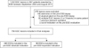
In this retrospective study, we reviewed the medical records of 967 consecutive patients who underwent ESD for 1,032 EGC lesions between September 2004 and August 2011 at the National Cancer Center, Korea. Of these EGC lesions, 276 were excluded because of the following: 1) were not adenocarcinomas as determined after a diagnostic endoscopic biopsy (adenoma lesions and atypical gland), 2) were diagnosed at the remnant stomach after subtotal gastrectomy, 3) were out-of-indication per the diagnostic evaluations, or 4) were multiple in same patients.
The baseline demographic characteristics; pre-ESD diagnostic findings, including endoscopy and pathological results; and final pathological evaluation results after ESD were obtained from the prospectively collected database. This study was approved by the Institutional Review Board (IRB) of the National Cancer Center, Korea (NCC2015-0059). Informed consent was waived for all the patients ts because the IRB assessed this sutdy as low-risk.
2. Definition of pre-endoscopic submucosal dissection (ESD) indications and pathological curability criteria for post-ESD indications
Based on pre-ESD evaluations and final pathological evaluation results after ESD, ESD indications were divided into the absolute and expanded indications according to the Japanese Gastric Cancer Treatment Guidelines.2 The pre-ESD depth of tumor invasion was clinically determined by using conventional white light endoscopy (WLE), abdominal computed tomography (CT), and/or endoscopic ultrasonography (EUS).
Pre-ESD absolute indication was defined as a differentiated tumor measuring ≤2 cm that has no ulcer and is confined to the mucosal layer.2 The pre-ESD expanded indications included the following types of tumors confined to the mucosal layer: 1) a differentiated tumor measuring >2 cm without an ulcer, 2) a differentiated tumor measuring ≤3 cm with an ulcer, and 3) an undifferentiated tumor measuring ≤2 cm without an ulcer.2 The presence of ulcer was defined when a definite visible ulcer was detected on the tumor upon endoscopic examination.
The final pathological curability criteria for absolute indications (post-ESD absolute criteria) and expanded indications (post-ESD expanded criteria) were defined as follows: The post-ESD absolute criteria were the same as the pre-ESD absolute indication criteria for the lesions without lymphovascular invasion. The post-ESD expanded criteria included the 3 aforementioned pre-ESD expanded indication criteria plus the criterion of a differentiated histological type with minute submucosal invasion (<500 µm) and a tumor size of ≤3 cm. All lesions that meet the post-ESD expanded criteria should have no lymphovascular invasion.2 Tumors out-of-indication were defined as tumors that did not meet the expanded criteria in the final pathological evaluations.
3. Pathological evaluation
The ESD procedures, described in a previous study,14 were performed by 4 experienced gastroenterologists. All the endoscopists were certified specialty board members of the Korean Society of Gastrointestinal Endoscopy and have performed more than 5,000 diagnostic endoscopic procedures. Resected ESD specimens were fixed in 10% formalin and then embedded in a paraffin block, which was serially sliced at 2-mm intervals. The paraffin slices were then stained with hematoxylin and eosin, and a single specialized pathologist (MC Kook) performed the pathological evaluation. The World Health Organization classification of gastric cancer was used for determination of tumor histological subtypes.15 Subsequently, well-differentiated and moderately differentiated tubular adenocarcinomas, as well as papillary adenocarcinoma, were included in the differentiated histological type. By contrast, poorly differentiated tubular adenocarcinoma, signet ring cell carcinoma, and mucinous adenocarcinoma were included in the undifferentiated histological type.2 Tumor histological types were determined according to the major component that constituted >50% of the tumor in cases of mixed histological types.2,15
4. Definitions of endoscopic submucosal dissection outcomes
En bloc resection was defined as removal of the tumor in one piece without fragmentation. Complete resection was defined as removal of the tumor using en bloc resection, with negative horizontal and vertical tumor resection margins.16 Curative resection was achieved when tumors were completely resected and final pathological evaluation results met the curability criteria for absolute or expanded indications of ER.2
5. Statistical analyses
Data were compared between the pre-ESD absolute and expanded indication groups by using the chi-square or Fisher exact test for categorical variables and the Student t-test or Mann-Whitney U-test for continuous variables. Univariate and multivariate logistic regression analyses were performed to investigate pre-ESD risk factors associated with out-of-indication that were identified upon final pathological evaluation after ESD. The covariates for the multivariate logistic regression analysis were pre-ESD variables that showed a statistical significance in the univariate analyses. P-values <0.05 were considered statistically significant. All data were analyzed by using Stata 12.1 (Stata, College Station, TX, USA).
Results
1. Baseline characteristics
Of the 967 EGC lesions, 756 were included in the final analyses (Fig. 1). These tumors were further classified into the pre-ESD absolute and expanded indication groups on the basis of diagnostic evaluations. EGC lesions met the pre-ESD absolute indications in 660 cases (87.3%) and the pre-ESD expanded indications in 96 cases (12.7%)
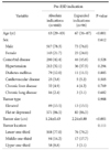
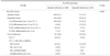
The median age of all the included patients was 64 years, and the proportion of male patients was 78.0%. Compared with the patients in the pre-ESD absolute indication group, those in the pre-ESD expanded indication group were significantly older (median age, 63 vs. 67 years; P<0.001) and had a larger mean tumor size (1.24 vs. 2.24 cm; P<0.001; Table 1). No significant differences in sex, comorbid disease, tumor type, and tumor location were observed between the two groups.
2. Short-term outcomes of endoscopic submucosal dissection (ESD) according to pre-ESD indication
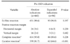
The en bloc resection rate in all the included lesions was 98.4%, and the rates did not different between the pre-ESD absolute and expanded indication groups (98.6% vs. 96.9%, respectively; P=0.197). The final pathological evaluation results of the resected specimens indicated no significant difference (P=0.228) in the complete resection rate between the pre-ESD absolute indication group (93.0%) and the pre-ESD expanded indication group (89.6%). However, the curative resection rate was significantly lower in the lesions with the pre-ESD expanded indications than in those with the pre-ESD absolute indications (64.6% vs. 81.7%, respectively; P<0.001; Table 2).
3. Discrepancies between pre-endoscopic submucosal dissection (ESD) and post-ESD criteria
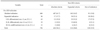
The discrepancies between the pre-ESD indications and post-ESD criteria are shown in Table 3. Overall, the final pathological evaluation results indicated that 15.9% of tumors met the out-of-indication criteria. Of the tumors in the pre-ESD absolute indication group, only 61.7% were correctly estimated according to the post-ESD pathological evaluation results, and the remaining 38.3% of the lesions were upgraded to either expanded indication (24.5%) or out-of-indication (13.8%). Of the tumors in the pre-ESD expanded indication group, only 52.1% were correctly est imated, and 30.2% met the out-of-indication criteria in the post-ESD pathological evaluation. Tumors with a pre-ESD expanded indication had a significantly higher rate of post-ESD out-of-indication than those with a pre-ESD absolute indication (13.8% vs. 30.2%, respectively; P<0.001). Among the three subgroups of pre-ESD expanded indications, the undifferentiated histological type subgroup had the highest rate of post-ESD out-of-indication (53.3%).
After ESD, the presence of lymphovascular and deep submucosal tumor invasions (≥500 µm) were the main causes of the out-of-indication in both pre-ESD indication groups. Details of the post-ESD criteria, determined via pathological evaluations, are compared with the pre-ESD indications in Table 4.
4. Pre-endoscopic submucosal dissection (ESD) characteristics associated with the post-ESD out-of-indication
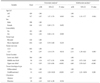
According to univariate analyses of the final pathological evaluation results, significant risk factors associated with out-of-indication include age of >65 years, tumor size of >2.0 cm, tumor in the upper-third segment of the stomach, and an undifferentiated histological type in the pre-ESD evaluations. Multivariate analysis confirmed that age of >65 years (adjusted odd ratio [aOR], 1.84; P=0.004), tumor size of >2.0 cm (aOR, 2.51; P=0.003), tumor in the upper-third segment of the stomach (aOR, 4.68; P<0.001), and an undifferentiated tumor (aOR, 6.47; P=0.001) were independent pre-ESD risk factors associated with out-of-indication (Table 5).
Discussion
EGC lesions that are estimated to meet the criteria for ESD indications are often found to be out-of-indication in post-ESD pathological evaluations. This situation results in the need for additional curative-intent surgery. In the present study, 15.9% of all patients who had met the pre-ESD indications for treatment in the diagnostic evaluations were identified as out-of-indication according to the post-ESD pathology results. Out-of-indication rates determined via final pathological evaluations were 30.2% in tumors that met the pre-ESD expanded indications and 13.8% in those that met pre-ESD absolute indications (P<0.001). The out-of-indication rate after ESD was highest (53.3%) in the tumors that met the pre-ESD expanded indications of undifferentiated tumors rs measuring ≤2 cm and wtihout an ulcer.
The ESD indication for EGC lesions were based on the negligible risks of lymph node metastasis that were derived from lymph node risk analysis in a large number of cases of surgically resected specimens.17 Tumor characteristics, including size, histological type, depth of invasion, and presence of ulceration are major factors to be considered in the estimation of lymph node metastasis risks.18 Clinical evaluations for determining the aforementioned factors before ESD include conventional WLE for tumor size estimation, ulcer findings, biopsy for tumor histology, and imaging studies (abdominal CT and/or EUS) for predicting depth of tumor invasion. However, current diagnostic modalities have limitations in accurate assessment, such as the underestimation of tumor size via conventional WLE19 and the limited efficacy of CT and EUS in predicting depth of tumor invasion.11,20,21 Hence, discrepancies between clinical indications of ESD and the final criteria for curative resection seems inevitable.
Previous studies that compared outcomes of ESD according to the post-ESD criteria reported that the en bloc resection rate did not differ between both post-ESD criteria, but complete resection rates were significantly lower in the expanded criteria than in the absolute criteria groups.3,9,10 In the present study, the en bloc and complete resection rates did not differ between both pre-ESD indication groups according to the pre-ESD evaluation results. As the endoscopists decided to perform ESD only for EGC lesions that met the ER indications in the pre-ESD evaluations, the complete resection rate did not differ between the expandedand absolute indication groups in the present study. However, the curative resection rate was significantly lower in the pre-ESD expanded indication group than in the pre-ESD absolute indication group (64.6% vs. 81.7%, respectively). In addition, a guideline states that ESD for EGC lesions that meet the expanded indication is defined as an investigational treatment.2 Therefore, caution should be exercised when choosing ESD as the primary treatment for patients with EGC lesions that meet the expanded indications in clinical evaluations, even though performing ESD for these lesions seems technically feasible.
In our study, the highest rate of post-ESD out-of-indication was observed for tumors classified under the pre-ESD expanded indication group of the undifferentiated histologic type subgroup (53.3%). This result is similar to those in previous studies that reported low curative resection rates (55%~65%) in patients who underwent ER for undifferentiated lesions.22,23,24 Many possible causes of noncurative resection after ESD for undifferentiated lesions have been suggested. First, tumor sizes tend to be larger than those estimated before ESD. For example, undifferentiated lesions have been reported to have significantly larger size discrepancies between pre-ESD and post-ESD evaluations than the differentiated lesions.20,25 Second, the submucosal tumor invasion could play a role in noncurative resection. In patients who undergo ESD for undifferentiated lesions, final pathological evaluations revealed submucosal invasion rates of 20% to 28%.13,23,25 In addition, previous studies reported significantly lower accuracies of pre-ESD prediction of depth of tumor invasion in lesions of the undifferentiated histologic type than in those of the differentiated histologic type.11 Magnifying endoscopy with narrow band imaging might be useful for accurate demarcation of undifferentiated EGC lesions.26 However, endoscopists should be careful in deciding whether to perform an ESD in patients with undifferentiated EGC.
In the present study, both lymphovascular and submucosal tumor invasions were the most common causes of out-of-indication. Of the components that comprised ER indications, diagnostic evaluations before ESD have limited roles in predicting the depth of tumor invasion and lymphovascular invasion. In predicting the depth of tumor invasion, the reported accuracy rates of pre-ESD evaluations are low, ranging from 63% to 74% as determined via conventional endoscopy11,27 and from 67% to 72% as determined via EUS.11,21,27,28 In addition, the presence of lymphovascular invasion is the single, most important risk factor of lymph node metastasis in EGC patients. However, this risk factor cannot be evaluated or predicted with pre-ESD evaluation; it can only be determined by performing a pathological evaluation of the resected specimen after ESD. These limitations of pre-ESD evaluations are the main reasons for the discrepancies between pre-ESD indication and post-ESD criteria. Further studies to investigate more-accurate methods for predicting the depth of tumor invasion and presence of lymphovascular invasion, if possible, are needed.
Previous studies reported that large tumor size, tumor location in the upper-third segment of the stomach, submucosal tumor invasion, and ulcer findings are the factors associated with noncurative resection after ESD.29,30,31 Similarly, the multivariate analysis in our study revealed that a tumor size of >2 cm, tumor location in the upper-third segment of the stomach, and an undifferentiated histologic type in the pre-ESD evaluations were independent risk factors. These results suggest that before ESD is performed, patients who have the aforementioned pre-ESD risk factors should be fully informed of the possibility of out-of-indication in the final pathological evaluation results and the possible need for additional surgical treatment.
An additional surgery is needed in patients with EGC lesions that are judged as out-of-indication after ESD.2 Patients who underwent noncurative resection after ESD who did not receive an additional surgical treatment had significantly lower OS than those who underwent an additional surgery.12,13 Despite this knowledge, a considerable number (44%~68%) of patients who undergo noncurative resection after ESD do not undergo additional surgery.4,10,12,13 Thus, endoscopists should carefully select candidates for ESD for EGC lesions based on precise pre-ESD diagnostic evaluations and explain to the patient that the expanded indications of ESD is for an investigational treatment.
The strength of the present study is that it investigated the discrepancies between pre-ESD indication and post-ESD criteria in a large number of EGC cases. In addition, pre-ESD risk factors of out-of-indication after ESD were analyzed. These results may be helpful for endoscopists to more precisely decide on ESD indications for EGC. The limitations of this study include the potential for selection bias caused by its retrospective design and single-center setting. Furthermore, not all patients underwent additional pre-ESD diagnostic evaluations by using narrow band imaging or EUS to estimate tumor size and depth of invasion. Another limitation is that the results of the pre-ESD evaluation to identify lesion characteristics might have slightly differed between endoscopists.
In conclusion, noncurative resection because of out-of-indication occurred in as many as one third of the cases of EGC lesions that clinically met the pre-ESD expanded indications. Therefore, the possibility of additional surgery should be emphasized to patients with these types of EGC lesions before they undergo ESD treatment, and minimally invasive surgery rather than ESD should be considered as the primary treatment for undifferentiated EGCs.
 XML Download