

 PDF
PDF ePub
ePub Citation
 Print
Print


Introduction
Helicobacter pylori infection is closely related to gastric atrophy, intestinal metaplasia (IM), and progression to dysplasia or cancer.1 However, the incidence of gastric cancer did not differ between Chinese patients receiving H. pylori eradication treatment and those receiving placebo during 7.5 years of follow-up.2 Several reports exist on the effect of H. pylori eradication in patients at a high risk for gastric cancer. Previous studies illustrated that eradication of H. pylori after endoscopic resection (ER) of early gastric cancer (EGC) helped prevent the development of metachronous gastric cancer.34 However, Choi et al.5 revealed that H. pylori eradication after ER of EGC did not reduce the incidence of metachronous gastric cancer. Therefore, the importance of H. pylori eradication after ER for gastric neoplasms remains controversial. Defining the subgroup of patients for whom H. pylori eradication after ER for gastric neoplasms is beneficial is important because patients who undergo ER is increasing and the incidence of metachronous lesions is also consequently increasing.
Several gastric and intestinal epithelial markers are associated with the progression of IM, a precancerous lesion.67 Sonic hedgehog (SHH) is an essential regulator of developmental and physiological processes.8910 In humans, Shh mRNA and SHH protein are expressed in the normal gastric fundus but not in the esophagus and intestine.1112 Loss of SHH is an early change associated with the presence of IM.1113 CDX proteins (intestine-specific caudal-related homeobox transcription factor) are intestine-specific transcription factors encoded by human CDX1 and CDX2 genes. Moreover, Mizoshita et al.14 reported that the expression of CDX1 and CDX2 was found in IM of the human stomach. Shiotani et al.7 demonstrated the reexpression of SHH and reduction of CDX2 levels after H. pylori eradication. Sox2, a gastric transcription factor, is expressed in the human stomach, and its expression gradually decreases as IM progresses.15 That is, Sox2 expression decreases and Cdx expression increases gradually during the progression of IM. Therefore, these markers might help identify patients for whom H. pylori eradication is advantageous. In this study, we evaluated the clinicopathological factors and gastric and intestinal markers according to the development of metachronous gastric neoplasms after ER, clarified the role of H. pylori eradication in the development of metachronous lesions, and investigated the patient subgroup for whom H. pylori eradication was beneficial, especially after ER.
Materials and Methods
1. Patients
Between June 2006 and June 2012, gastric neoplasms were diagnosed in 3,882 patients and treated with ER. Among the patients, we identified those who were infected with H. pylori and received H. pylori eradication treatment; that is, all patients included in this study received H. pylori eradication treatment. After the H. pylori eradication treatment, 34 patients with metachronous gastric neoplasms after ER for gastric neoplasms (metachronous group) and 102 age- and sex-matched patients (nonmetachronous) who underwent ER for gastric neoplasms and who did not develop metachronous gastric neoplasms were enrolled on the basis of propensity score matching at a ratio of 1:3. The metachronous and nonmetachronous groups were matched according to age and sex by using propensity score matching. We defined metachronous lesions as those that developed at another site (not a previous ER site) at 6 months or later after the previous ER. The period between the development of metachronous gastric neoplasms and H. pylori eradication was at least 6 months, to accurately evaluate the effect of H. pylori eradication on the incidence of metachronous lesions after ER.4
In addition, there was a median interval of 3 months (range, 0~32 months) between ER and H. pylori eradication. To determine the H. pylori infection status, rapid urease and urea breath tests and histological assessment were done. Patients who had a positive result for at least one of these tests were defined as the group with a positive H. pylori infection. In contrast, those who showed negative results for all three examinations were defined as the group with negative H. pylori infection. The first eradication regimen was conventional triple therapy consisting of standard doses of a proton pump inhibitor (PPI) twice daily, 1,000 mg amoxicillin twice daily, and 500 mg clarithromycin twice daily for 7 to 14 days. In addition, in cases of eradication failure with the initial conventional triple therapy, quadruple therapy including two standard doses of a PPI, 500 mg metronidazole thrice daily, 120 mg bismuth four times daily, and 500 mg tetracycline four times daily for 7 to 14 days was used. The patients were evaluated in terms of their clinicopathological characteristics, such as age, sex, tumor location, gross appearance of the tumor, tumor histological type, tumor size, tumor invasion depth, tumor multiplicity, the presence of ulcers, background mucosa, and H. pylori eradication failure rate. Atrophy and IM were evaluated endoscopically at the time of ER. In particular, atrophic gastric mucosa was categorized by using the Kimura classification.1617 The severity of atrophy was categorized as open or closed, and that of IM was classified as none, mild, moderate, or severe. Mild grade was characterized as metaplastic mucosa with fine or granular plaques. Moderate grade was defined as coarse plaques or patches. Severe grade was characterized as mucosa with coarser and larger plaques or patches.17 H. pylori infection was evaluated via histopathological examinations of the resected specimen, the urea breath test, or the rapid urease test. The clinicopathological characteristics were analyzed retrospectively and compared between the metachronous and nonmetachronous groups. The median period between the initial resection and the subsequent identification of metachronous neoplasm was 30.1 months (range, 9.5~74.1 months), and that between the initial resection and the determination of no neoplasm (nonmetachronous group) was 49.1 months (range, 16.7~92.7 months). We excluded metachronous recurrence occurring within 6 months because the lesions detected during this time were most likely lesions that were missed before and that should be categorizeads synchronous cancers or neoplasms.
The Institutional Review Board of Severance and Gangnam Severance Hospital approved this study (3-2014-0117).
2. Immunohistochemistry
Immunohistochemical staining was performed for SHH, CDX1, CDX2, and SOX2. Twenty patients were selected for immunohistochemistry on the basis of the development of metachronous neoplasms before and after H. pylori eradication, with 10 specimens obtained from each group both before and after H. pylori eradication. Immunohistochemical staining was performed after H. pylori eradication at the time of the subsequent identification of metachronous neoplasm (metachronous group, median 30.1 months) and the determination of no neoplasm (nonmetachronous group, median 49.1 months). Biopsy specimens were obtained from the antrum and corpus for evaluating the background mucosa by using immunohistochemical staining. We used paraffin-embedded tissue specimens and rabbit polyclonal anti-SHH (Santa Cruz Biotechnology, Santa Cruz, CA, USA), goat polyclonal anti-SOX2 (Santa Cruz Biotechnology), mouse monoclonal anti-CDX1 (Santa Cruz Biotechnology), and rabbit polyclonal anti-CDX2 (Santa Cruz Biotechnology) antibodies. The expression levels were determined by scoring both the positivity rate and intensity of SHH, CDX1, CDX2, and SOX2 staining. The positive expression rate was determined by counting the number of immunopositive cells in three areas randomly chosen in the tissue for a total of 300 cells and scored (0=0%, 1=1%~25%, 2=26%~35%, 3=36%~45%, 4=46%~65%, 5=66%~100%). The staining intensity was scored as follows: 0, no detectable cytoplasmic staining; 1, mild cytoplasmic staining; 2, moderate cytoplasmic staining; and 3, severe cytoplasmic staining. The two scores were then summed to obtain a final score between 0 and 8.
3. Statistical analysis
The chi-square test and Fisher exact test were used to compare the clinicopathological factors between the groups according to the development of metachronous gastric neoplasms. The t-test was used for noncategorical variables in the intergroup comparisons of clinicopathological characteristics. The Wilcoxon signed-rank test and Mann-Whitney U-test were used for evaluating immunohistochemical expression levels. The metachronous and nonmetachronous groups were matched according to age and sex by using propensity score matching at a ratio 1o:f3. Statistical significance was set at P<0.05. All statistical analyses were performed by using SPSS ver. 12.0 for Windows (SPSS Inc., Chicago, IL, USA).
Results
1. Clinicopathological characteristics of the metachronous and nonmetachronous groups
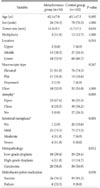
The clinicopathological features of 34 patients with metachronous gastric neoplasms and 102 nonmetachronous patients are shown in Table 1. No differences were found in tumor size, multiplicity, location, macroscopic type, ulcer, and histology between the metachronous and nonmetachronous groups. Open-type atrophy (P=0.003), moderate and severe IM (P=0.001), and failure of H. pylori eradication (P=0.036) occurred more frequently in the metachronous group (Table 1).
2. Demographic and clinicopathological characteristics of the metachronous and nonmetachronous groups according to histopathology
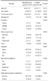
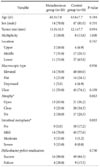
Of the 136 patients who underwent ER for gastric neoplasms, 58 (42.6%) underwent ER for dysplasia and 78 (57.4%) underwent ER for carcinoma. In patients with dysplasia, endoscopically flat-type lesions (P=0.030), ulcers (P=0.043), and open-type atrophy (P=0.047) were found predominantly in the metachronous group. The failure rate of H. pylori eradication was significantly higher in the metachronous group than in the nonmetachronous group (P=0.002) (Table 2). In patients with carcinoma, moderate-to-severe IM was observed more frequently in the metachronous group (P=0.003). The failure rate of H. pylori eradication was not different between the metachronous and nonmetachronous groups (P=0.730) (Table 3).
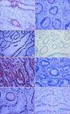
3. Immunohistochemistry
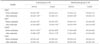
To investigate gastric and intestinal markers according to the development of metachronous gastric neoplasms after ER, immunohistochemical staining was performed for 10 metachronou s and 10 nonmetachronous patients before and after H. pylori eradication (Fig. 1). The intensities of SOX2 staining in the antrum and SHH staining in the antrum and corpus were increased in the nonmetachronous group after H. pylori eradication (P<0.05). In addition, the intensity of CDX2 staining in the antrum and corpus was decreased in the nonmetachronous group after H. pylori eradication (P<0.05). However, such changes in the intensity of SOX2, SHH, and CDX2 staining were not observed in the metachronous group. The intensity of SOX2 and SHH staining in the antrum after H. pylori eradication was significantly lower in the metachronous group than in the nonmetachronous group (P<0.05). Moreover, the intensity of CDX2 staining in the antrum before H. pylori eradication was significantly higher in the metachronous group than in the nonmetachronous group (P<0.0 5) (Table 4).
Discussion
The 5-year survival rate of EGC was favorable (>95%).18 ER is currently accepted as a local treatment for EGC, and the long-term outcome of ER for EGC in Korea is favorable.1920 ER has the advantages of preserving the entire stomach and increasing the postoperative quality of life of patients; however, the risk of synchronous or metachronous gastric cancer may be higher in the remnant stomach compared with that after gastrectomy. Metachronous gastric cancer developed in 2.7% to 14.0% of patients after ER within 3 to 5 years.2122 Multiple gastric lesions are more frequent in EGC than in advanced gastric cancer. Therefore, the detection of metachronous lesions after ER is important during follow-up after ER. In addition, identifying patients at a high risk of developing metachronous lesions and investigating the clinicopathological factors associated with metachronous lesions are clinically important.
Chronic H. pylori infection is associated with gastric cancer; however, the effect of H. pylori eradication on the prevention of metachronous gastric cancers remains controversial. Choi et al.5 reported that H. pylori eradication after ER of gastric tumor did not reduce the incidence of metachronous gastric carcinoma. In this study, persistent H. pylori infection was found in 80 of 439 patients who underwent H. pylori eradication therapy. Moreover, metachronous gastric cancer developed in 10 patients in whom H. pylori had been eradicated (10 of 439, 2.3%). According to histology, metachronous gastric cancer developed in three patients with dysplasia (3 of 226, 1.3%) and seven patients with carcinoma (7 of 213, 3.3%) after H. pylori eradication. Kwon et al. reported that persistent H. pylori infection was significantly associated with metachronous gastric cancer. The incidence of metachronous gastric lesions was significantly higher in patients with persistent H. pylori infection than in those in whom the bacterium was eradicated (18.8% vs. 8.4%, P=0.016).23
In the present study, all patients were infected with H. pylori and received H. pylori eradication treatment. We selected such a subgroup because we wanted to identify the patients for whom H. pylori eradication treatment would be helpful. Previous studies35 reported controversial results about the effects of H. pylori eradication treatment on metachronous gastric neoplasms. Therefore, we selected patients who received H. pylori eradication treatment, and then investigated the clinicopathological factors that affected the development of metachronous neoplasms. In this study, the number of patients with metachronous gastric neoplasms after ER was 34 (14 with dysplasia, 20 with carcinoma). However, the number of patients with metachronous gastric neoplasms was not small. Similar to previous studies, metachronous recurrence was observed in 33 patients during 3 years of follow-up3 and in 27 patients during 3 years of follow-up.5 Although the 3,882 patients were found to have gastric neoplasms and were scheduled for ER, the final group for the study consisted of 136 patients because we focused on those patients who received H. pylori eradication treatment. In addition, we investigated the role of H. pylori infection on the development of metachronous lesions by comparing the failure rate of H. pylori eradication; that is, we assessed the effect of persistent H. pylori infection on metachronous lesions after ER. Open-type atrophy, severe IM, and failure of H. pylori eradication were significantly associated with metachronous gastric neoplasms. Notably, failure of H. pylori eradication was significantly more frequent in metachronous patients with gastric dysplasia but not those with carcinoma.
McDonald et al. reported that genetic/epigenetic changes in IM would lead to dysplasia by identifying identical genetic alterations in dysplastic lesions and the surrounding IM; indeed, gastric cancer can arise from dysplastic lesions through further genetic alterations, referred to as 'cancer in adenoma'.2425 The causes of genetic and epigenetic changes in IM or dysplasia are diverse, and several genetic and epigenetic changes were reversed by H. pylori eradication. In this study, persistent H. pylori infection was significantly more frequent in patients with gastric dysplasia but not in those with carcinoma, in the metachronous group than in the nonmetachronous group. Therefore, H. pylori infection may affect the development of metachronous gastric neoplasms in patients with dysplasia or precancerous lesions before carcinomatous transformation. Only moderate-to-severe IM was significantly associated with carcinoma in the metachronous group. The reason might be the 'point of no return' the point at which H. pylori eradication could no longer prevent metachronous lesions. Accurately defining the point of no return is difficult because the molecular process cannot be measured precisely. However, our data suggest that H. pylori eradication may be more effective before the status of the background mucosa progresses to severe IM.
Currently, H. pylori eradication for preventing metachronous gastric carcinoma after ER is permitted but is not covered by the Korean National Health Insurance. Meanwhile, H. pylori eradication is not covered for precancerous lesions such as atrophy, IM, or dysplasia because of the lack of sufficient data. However, in this study, the role of H. pylori eradication in the prevention of metachronous lesions was more important in dysplasia than in carcinoma. Therefore, H. pylori eradication would be more helpful in patients with dysplasia than in those with carcinoma after ER. Previous reports about the association between H. pylori eradication and metachronous lesions have focused on EGC. However, on the basis of our results, H. pylori eradication after ER should be performed in patients with dysplasia for the prevention of metachronous lesions.
Our study has some limitations. This study was retrospective, and H. pylori eradication treatment could not be randomized. In addition, the period between ER and H. pylori eradication varied. The background mucosa such as atrophy and IM were evaluated endoscopically, not histologically.
Bornschein et al.26 reported that the expression levels of CDX1, CDX2, and SOX2 were similar in gastric cancer and in the surrounding nontumorous mucosa. They compared the gene expression between the tumor and the tumor-distant mucosa. In addition, upregulation of CDX1 and CDX2 and downregulation of SOX2 in the nonneoplastic gastric mucosa of patients with gastric cancer were confirmed. Moreover, H. pylori infection had no significant effect on the regulation of CDX1, CDX2, and SOX2 at the gastric cancer stage. In this study, the expression of CDX2 decreased and that of SOX2 increased after H. pylori eradication in the nonmetachronous group in which metachronous gastric neoplasm did not arise. The transgenic expressiono f cdx2 in the murine stomach can induce IM, which progresses to invasive gastric cancer.27 Furthermore, SOX2 is downregulated in gastric carcinoma.1528 Therefore, the negative correlation of CDX2 and inverse relation with SOX2 expression in this study might be associated with the effects of H. pylori eradication on premalignant lesions before carcinoma. SHH expression also increased after H. pylori eradication in the nonmetachronous group. Shiotani et al.7 reported that H. pylori eradication was associated with an increase in SHH expression in the corpus, especially in the absence of incomplete IM. Incomplete IM at the corpus is more common in patients with gastric cancer,13 which is in agreement with our finding of a positive correlation with SHH expression in the nonmetachronous group after H. pylori eradication. In summary, the expression of SHH and SOX2 increased and that of CDX2 decreased after H. pylori eradication in the nonmetachronous group in this study. Although the results were derived from a small sample size, these markers could be used to define the subgroup in which H. pylori eradication would be beneficial after ER.
In conclusion, H. pylori eradication can play an important role in the prevention of metachronous lesions after ER in patients with precancerous lesions before carcinomatous transformation. In addition, the changes in SHH, SOX2, and CDX2 expression after H. pylori eradication may be predictive markers for metachronous lesions after ER.
 XML Download