Abstract
Summary of Literature Review
Osteochondroma is one of the most common benign tumor in bone, consist of 40%, but, rare in spine area occupying only 2%. We report a case of osteochondroma that was in the 12th vertebra of thoracic spine, that had severe right flank pain. We performed en bloc excisional biopsy of the bony mass.
Materials and Methods
A fourty seven-year-old male complained right flank pain. He had mass of 12th thoracic costovertebral junction and underwent open excision and biopsy
REFERENCES
2. Yagi M, Ninomiya K, Kihara M, Horiuchi Y. Symptomatic osteochondroma of the spine in elderly patients. J Neurosurg Spine. 2009; 11:64–70.

3. Cooke RS, Cumming WJ, Cowie RA. Osteochondroma of the cervical spine: case report and review of the literature. Br J Neurosurg. 1994; 8:359–63.

4. Chung SS, Lee JS, Kim DJ, Moon SH, Yoon TH. Osteochondroma of the Cervical Spine: A Case Report. J Korean Orthop Assoc. 2004; 39:833–6.

6. Dahlin DC, Unni KK. Bone Tumors: General Aspects and Data on 11,087 cases. 5th ed.Springfield, IL: Lippincott-Raven;1996. 1:p. 11–25.
7. Malat I, Virapongse C, Levine A. Solitary osteochondroma of the spine. Spine (Phila Pa 1976). 1986; 11:625–8.

8. Huvos AG. Bone tumors, Diagnosis Treatment and Prognosis. 2nd ed.Philadelphia: W.B. Ssunders Co.;1991.
9. MacGee EE. Osteochondroma of the cervical spine: a cause of transient quadriplegia. Neurosurgery. 1979; 4:259–60.
10. Madigan R, Worrall T, McClain EJ. Cervical cord compression in hereditary multiple exostosis: Review of the literature and report of a case. J Bone Joint Surg Am. 1974; 56:401–4.
Fig. 1.
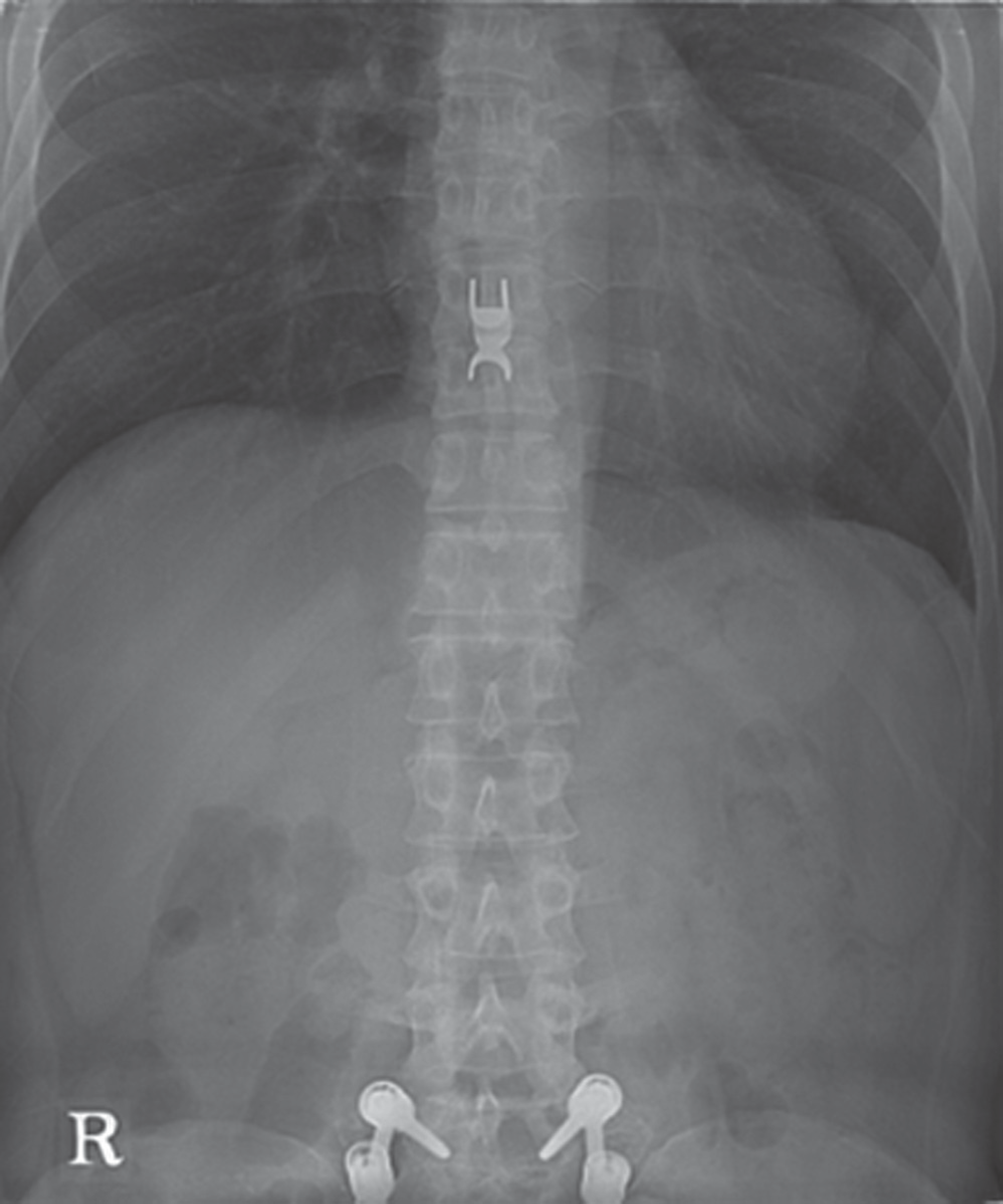
(A) Four years ago, anteroposterior radiography of lumbar spine of 47 years old man showed that 12th costovertebral junction was clear. (B) Bony mass which looks radioopeque is found on 12th costovertebral junction in pre operative state. Radiographs of the thoracic spine, anteroposterior view show sclerotic and hypertrophic bony mass density on the costovetebral junction of the 12 th thoracic vertebrae.

Fig. 2.
(A) Axial computed tomography scan of the patient shows the osteochondroma protruding posteriorlaterally from the costovertebral junction of 12 th thoracic vertebra. We can find more hyperplastic costovertebral junction in Rt side than Lt which is normal. (B) MRI image. T1 weighted image. (C) T2 weighted image. In the thoracic MRI, it shows a irregular margined, well-circumscribed ossified mass lesion, from right side of 12 th thoracic vertebra on (B) T1 axial image and (C) T2 axial image.

Fig. 3.
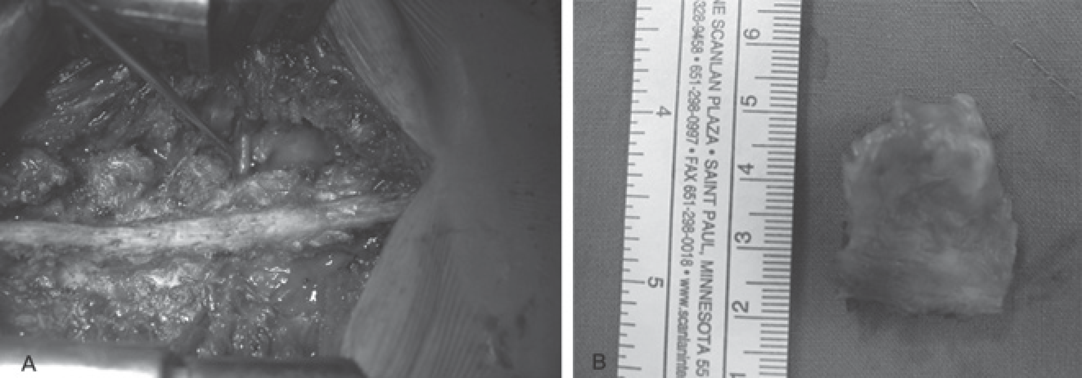
(A) Intra operative photography shows that 12 th thoracic nerve root is decompressed by excision of the mass occupied costovertebral junction space. (B) 3 X 2 X 2 cm sized oval shaped, irregular surfaced osteochondroma protruding posteriorly from the 12th thoracic vertebrae is removed.


 PDF
PDF ePub
ePub Citation
 Print
Print


 XML Download