Abstract
Objectives
We wanted to evaluate the clinical and radiological outcomes of operative treatment for lumbar degenerative diseases in patients who are undergoing dialysis.
Summary of the Literature Review
Operative treatment for patients having spinal diseases with chronic renal failure (CRF) demands special consideration because of the medical and surgical complications and the poor clinical outcome. There are only few reports on operative treatment for lumbar degenerative diseases for patients who are undergoing dialysis.
Materials and Methods
Eight patients with lumbar degenerative spondylopathy and CRF and who were undergoing dialysis were operated on from August 1998 to September 2007. The clinical and radiological outcomes were evaluated using the Japanese Orthopaedic Association (JOA) scale and the plain X-rays. The serum alkaline phosphatase levels were measured to evaluate the bone metabolism along with the postoperative improvement of clinical symptom.
Results
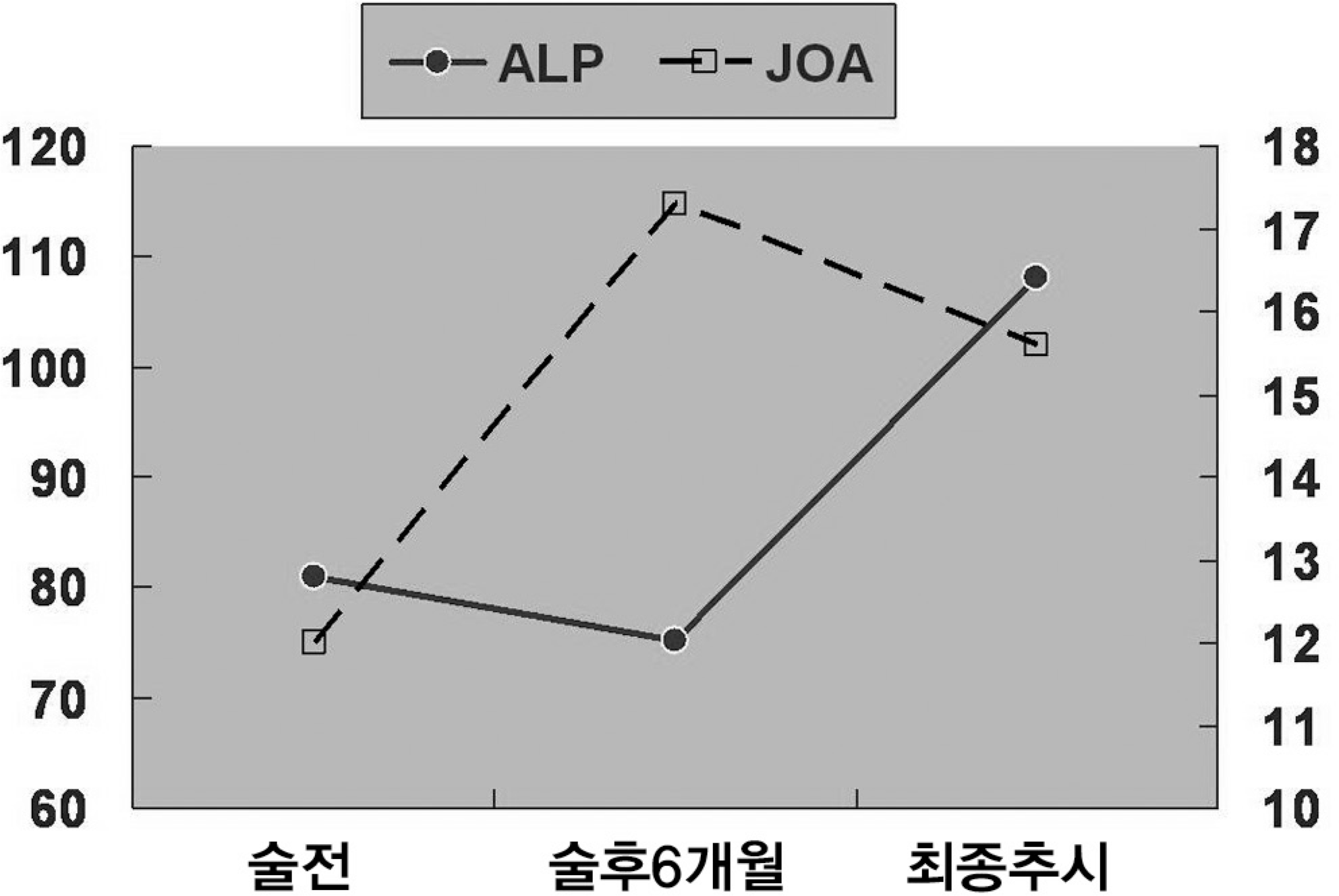
We had 1 case of postoperative mortality with peritoneal dialysis due to sepsis that was caused by panperitonitis and another complication of discitis. Only 1 of 4 cases that underwent fusion procedure had radiological bony union. The mean JOA scores were 12.0 (range: 10-14) preoperatively and 17.3 (range: 5-20) and 15.6 (range: 9-19) at postoperative 6 months and the final follow-up, respectively (p<0.05). The mean serum alkaline phosphatase levels were 80.9 (range: 43-142) preoperatively, 98 (range: 68-164) at postoperative 1 month, 75 (range: 50-102) at postoperative 6 months and 108 (range: 60-209) at the final follow-up (p>0.05).
Conclusions
The clinical outcomes of surgical treatments were improved for the degenerative spine disease patients who are undergoing dialysis. However after the fusion procedure, the bony fusion rate was low (25%). Since a high rate of perioperative medical complications can be expected, thorough medical evaluation during preoperation and postoperation is recommended.
REFERENCES
1.Marcelli C., Perennou D., Cyteval C, et al. Amyloidosis-related cauda equina compression in long-term hemodialysis patients. Three case reports. Spine. 1996. 21:381–5.
2.Kumar A., Leventhal MR., Freedman EL., Coburn J., Delamarter R. Destructive spondyloarthropathy of the cervical spine in patients with chronic renal failure. Spine. 1997. 22:573–7.

3.Maruyama H., Gejyo F., Arakawa M. Clinical studies of destructive spondyloarthropathy in long-term hemodialysis patients. Nephron. 1992. 61:37–44.

4.Christensen FB., Laursen M., Gelineck J., Eiskjaer SP., Thomsen K., Bunger CE. Interobserver and intraobserver agreement of radiograph interpretation with and without pedicle screw implants: the need for a detailed classification system in posterolateral spinal fusion. Spine. 2001. 26:538–43.
5.Sudo H., Ito M., Abumi K, et al. Long-term follow up of surgical outcomes in patients with cervical disorders undergoing hemodialysis. J Neurosurg Spine. 2006. 5:313–9.

6.Kuntz D., Naveau B., Bardin T., Drueke T., Treves R., Dryll A. Destructive spondylarthropathy in hemodialyzed patients. A new syndrome. Arthritis Rheum. 1984. 27:369–75.
7.Allain TJ., Stevens PE., Bridges LR., Phillips ME. Dialysis myelopathy: quadriparesis due to extradural amyloid of beta 2 microglobulin origin. Br Med J. 1988. 296:752–3.
8.Rousselin B., Helenon O., Zingraff J, et al. Pseudotumor of the craniocervical junction during long-term hemodialysis. Arthritis Rheum. 1990. 33:1567–73.

9.Ohashi K., Hara M., Kawai R, et al. Cervical discs are most susceptible to beta 2-microglobulin amyloid deposition in the vertebral column. Kidney Int. 1992. 41:1646–52.
10.Orzincolo C., Bedani PL., Scutellari PN., Ghedini M., Cardona P. [Course of radiologic changes in spondyloarthropathy caused by dialysis]. Radiol Med. 1991. 81:228–33.
11.Orzincolo C., Bedani PL., Scutellari PN., Cardona P., Trotta F., Gilli P. Destructive spondyloarthropathy and radiographic follow-up in hemodialysis patients. Skeletal Radiol. 1990. 19:483–7.

12.Bindi P., Lavaud S., Bernieh B., Toupance O., Chanard J. Early and late occurrences of destructive spondyloarthropathy in haemodialysed patients. Nephrol Dial Transplant. 1990. 5:199–203.

13.Otsubo S., Kimata N., Okutsu I, et al. Characteristics of dialysis-related amyloidosis in patients on haemodialysis therapy for more than 30 years. Nephrol Dial Transplant. 2009. 24:1593–8.

14.Cianciolo G., Coli L., La Manna G, et al. Is beta2-microglobulin-related amyloidosis of hemodialysis patients a multifactorial disease? A new pathogenetic approach. Int J Artif Organs. 2007. 30:864–78.
15.Leone A., Sundaram M., Cerase A., Magnavita N., Tazza L., Marano P. Destructive spondyloarthropathy of the cervical spine in long-term hemodialyzed patients: a five-year clinical radiological prospective study. Skeletal Radiol. 2001. 30:431–41.

16.Jadoul M., Garbar C., Noel H, et al. Histological prevalence of beta 2-microglobulin amyloidosis in hemodialysis: a prospective post-mortem study. Kidney Int. 1997. 51:1928–32.
17.Yuzawa Y., Kamimura M., Nakagawa H, et al. Surgical treatment with instrumentation for severely destructive spondyloarthropathy of cervical spine. J Spinal Disord Tech. 2005. 18:23–8.

18.Abumi K., Ito M., Kaneda K. Surgical treatment of cervical destructive spondyloarthropathy(DSA). Spine. 2000. 25:2899–905.
19.Nair S., Vender J., McCormack TM., Black P. Renal osteodystrophy of the cervical spine: neurosurgical implications. Neurosurgery. 1993. 33:349–54.
20.Sasaki M., Abekura M., Morris S, et al. Microscopic bilateral decompression through unilateral laminotomy for lumbar canal stenosis in patients undergoing hemodialysis. J Neurosurg Spine. 2006. 5:494–9.

21.Yamamoto T., Matsuyama Y., Tsuji T., Nakamura H., Yanase M., Ishiguro N. Destructive spondyloarthropathy in hemodialysis patients: comparison between patients with and those without destructive spondyloarthropathy. J Spinal Disord Tech. 2005. 18:283–5.
22.Van Driessche S., Goutallier D., Odent T, et al. Surgical treatment of destructive cervical spondyloarthropathy with neurologic impairment in hemodialysis patients. Spine. 2006. 31:705–11.

23.Shiota E., Naito M., Tsuchiya K. Surgical therapy for dialysis-related spondyloarthropathy: review of 30 cases. J Spinal Disord. 2001. 14:165–71.

24.Ersoy FF. Osteoporosis in the elderly with chronic kidney disease. Int Urol Nephrol. 2007. 39:321–31.

25.Han IH., Kim KS., Park HC, et al. Spinal surgery in patients with end-stage renal disease undergoing hemodialysis therapy. Spine. 2009. 34:1990–4.

26.Muraki S., Yamamoto S., Ishibashi H, et al. Impact of degenerative spinal diseases on bone mineral density of the lumbar spine in elderly women. Osteoporos Int. 2004. 15:724–8.

27.Erlichman M., Holohan TV. Bone densitometry: patients with end-stage renal disease. Health Technol Assess. 1996. 8:1–27.
28.Sit D., Kadiroglu AK., Kayabasi H., Atay AE., Yilmaz Z., Yilmaz ME. Relationship between bone mineral density and biochemical markers of bone turnover in hemodialysis patients. Adv Ther. 2007. 24:987–95.

29.Kim HJ., Lee HM., Kim HS, et al. Bone metabolism in postmenopausal women with lumbar spinal stenosis: analysis of bone mineral density and bone turnover markers. Spine. 2008. 33:2435–9.
Fig. 1 .
The graph shows the serial changes of JOA score and alkaline phosphatase levels with follow-up. No statistical correlation between them was observed with multiple regression analysis(p>0.05).

Table 1.
Summary of cases
Table 2.
Summary of the JOA scoring system, excluding bladder function∗
| Items | Score |
|---|---|
| Subjective symptoms (9 points) | |
| Low-back pain | |
| none | 3 |
| occasionally mild | 2 |
| always present or sometimes severe | 1 |
| always severe | 0 |
| Leg pain &/or numbness | |
| none | 3 |
| occasionally mild | 2 |
| always present or sometimes severe | 1 |
| always severe | 0 |
| Walking ability | |
| normal | 3 |
| able to walk .500 m, w/ pain/numbness/weakness | 2 |
| present | 1 |
| unable to walk 500 m due to pain/numbness/weakness | 0 |
| unable to walk 100 m due to pain/numbness/weakness | 2 |
| Objective signs (6 points) | 1 |
| SLR | 0 |
| normal | |
| 30–70° | 2 |
| <30° | 1 |
| Sensory function | 0 |
| normal | |
| mild disturbance | 2 |
| mild disturbance | 2 |
| apparent disturbance | 1 |
| Motor function | 0 |
| normal (MMT normal) | |
| slightly decreased muscle strength (MMT good) | 2 |
| marked decreased muscle strength (MMT, fair) | 1 |
| Restriction of ADL (14 points)† | 0 |
| none | |
| moderate | |
| severe | |
| Total score | 29 |
Table 3.
Operative treatment and results


 PDF
PDF ePub
ePub Citation
 Print
Print


 XML Download