

 PDF
PDF ePub
ePub Citation
 Print
Print


Introduction
Variations of the brachial plexus regarding the composition of nerve fibres and absence or communications are common and being reported by several authors [1, 2]. The median nerve is formed anterior or anterolateral to axillary artery by the union of its two roots. The lateral root of median nerve is the largest branch of the lateral cord of brachial plexus while the medial root arises from the medial cord (C8, T1) and crosses in front of the axillary artery to join the lateral root of the lateral cord (C5, 6, 7). After joining of both roots the median nerve descends anterior to the axillary artery and upper part of brachial artery to reach the medial aspect of brachial artery in the distal half of the arm. It gives off vascular branches to the brachial artery and usually a branch to pronator teres, a variable distance proximal to the elbow joint but no cutaneous branch in the arm.
The median nerve usually enters the forearm between the heads of pronator teres. It is separated from ulnar artery by the deep head of pronator teres. It passes behind a tendinous bridge between the humero-ulnar and radial heads of the flexor digitorum superficialis. Then descends posterior to flexor digitorum superficialis and anterior to flexor digitorum profundus. About 5 cm proximal to the flexor retinaculum it emerges from behind the lateral edge of flexor digitorum superficialis, and becomes superficial just proximal to the wrist. Here it lies between the tendons of flexor digitorum superficialis and flexor carpi radialis. It then passes deep to the flexor retinaculum into the palm. In the forearm it gives the anterior interosseous nerve, muscular branches, articular branches, the palmar cutaneous branch [3]. The anatomical variations of peripheral nerves are important to the neurosurgeons, orthopedic surgeons, neurologists and anatomists.
Case Report
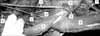
During educational gross anatomy dissection of embalmed 55-year-old male cadaver in the Department of Anatomy, All India Institute of Medical Sciences, New Delhi, we found, a variation in the right arm and forearm. On exploring the passage of median nerve in the upper part of the right arm it was found that an anomalous branch arising from the posterior aspect of the median nerve (Fig. 1) which was passing deep to the tendon biceps brachii (Fig. 2). Later it enters the cubital fossa where it is accompanied by the superficial vein of the forearm and then it terminates in the mid of the front of the forearm. The superficial vein was draining into the venae comitantes of the brachial artery in the cubital fossa (Fig. 2). The length of the anomalous cutaneous branch was about 18-20 cm.
The medial and lateral cutaneous nerves of the forearm were found to be normal. There were no communications between the branches of the cords. The relations of all the three cords of brachial plexus with the second part of axillary artery and the further course, branching and termination of the musculocutaneous nerve, median nerve, ulnar nerve and radial nerve in arm, forearm and hand followed the normal pattern. The other limb of the cadaver did not show any such variation and was absolutely normal in relation, formation and branching pattern of brachial plexus. No other neural, arterial or muscular variation was observed in either of the limbs.
Discussion
In this case report, flexure muscles of the arm were found to be innervated by the musculocutaneous nerve. An anomalous cutaneous branch arising from the median nerve in the right arm which was passing deep to the tendon of the biceps brachii. Later it enters the cubital fossa and then it becomes cutaneous which is accompanied by the superficial vein of the forearm. The median nerve supplying the flexure muscles of the forearm except the flexor carpi ulnaris and medial half of flexor digitorum profundus. In present case, there was no communication found between the musculocutaneous nerve and the median nerve as described by some authors [4].
The presence of this variation described in this case report may be because of the random factors influencing the mechanism of formation of the peripheral nerves during the embryonic life. In the humans, the forelimb muscles develop from the mesenchyme of paraxial mesoderm in the fifth week of the intrauterine life. The regional expression of five Hox D (Hox D 1 to Hox D 5) genes is responsible for upper limb development [5]. The guidance of the developing axons is regulated by the expression of chemoattractants and chemorepulsant in a highly coordinated site specific fashion. Tropic substances such as the brain-derived neurotropic growth factor, c-kit ligand, neutrin-1, neutrin-2, etc. attract the correct growth cones or support the viability of the growth cones that happen to take the right path [6]. The variations in nerve pattern may be the consequence of altered signaling between the mesenchymal cells and the neuronal growth cones or circulatory factors.
The brachial plexus anatomy variations are common and have been well documented in literature. Unilateral brachial plexus anomalies were demonstrated in 4 of 71 cadavers by Sarsilmaz et al. [7]. In present case report, we have also found that the variation was only in the right arm.
An atypical variant of the brachial plexus is the absence of the musculocutaneous nerve and innervation of the anterior compartment muscles of the upper arm including brachialis by the median nerve is also documented [2]. The branch to the brachialis muscle from the median nerve continued as the lateral cutaneous nerve of the forearm as described by Yogesh et al. in 2010 [8]. Beheiry EE in 2004 also reported that in 1.7% cases the median nerve gave muscular branches to the brachialis as well as a branch from its lateral root to supply both heads of the biceps brachii muscle in the absence of musculocutaneous nerve [9]. Contrast to the previous studies, in the present case, the median nerve was giving only cutaneous branch in the right arm to the front of the forearm.
Clinical significance
Theoretically, in a subject with an anomalous branch that we have described, injury will not result in loss of motor function but variable area of anesthesia or hypoesthesia may be produced. Cutaneous nerve in proximity to the superficial vein can be inadvertently damaged during venepuncture and may be a cause of pain following such procedures [10]. The length of the anomalous branch in our case was about 18-20 cm. The incidence of this anomalous cutaneous branch of the median nerve is not known, but its lengthy course makes it available for the functional restoration of irreparable lesions of the peripheral nerves. Consideration of possible anatomical variations of the median nerve is important when approaching the upper limb surgically and during trauma. Meticulous knowledge of such anatomical variations is important in interpreting atypical clinical presentations.

 XML Download