

 PDF
PDF ePub
ePub Citation
 Print
Print


Abstract
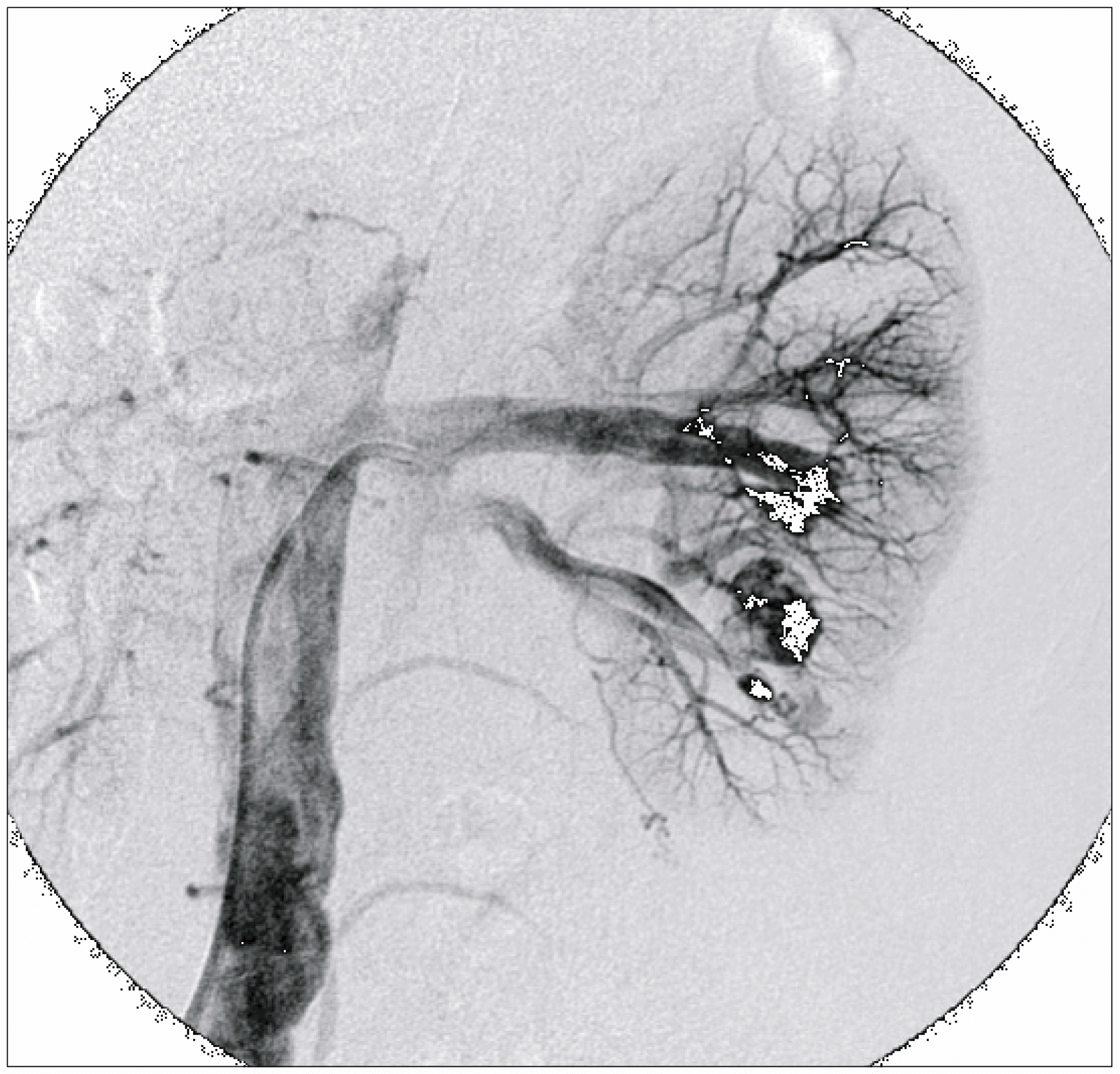
Currently, partial nephrectomy for patients with malignant renal tumors has become the procedure of choice for elective indications. Attempts have been made to use minimally invasive endoscopic procedures to replace the standard open partial nephrectomy. Laparoscopic partial nephrectomy can be technically challenging and be associated with vascular complications such as pseudoaneurysm. We report here on a case of renal artery pseudoaneurysm that occurred after laproscopic partial nephrectomy for renal cell carcinoma treated by percutaneous selective angioembolization.
References
1. Swana HS, Cohn SM, Burns GA, Egglin TK. Renal artery pseudoaneurysm after blunt abdominal trauma: case report and literature review. J Trauma. 1996; 40:459–61.
2. Dodd GD III, Tublin ME, Shah A, Zajko AB. Imaging of vascular complications associated with renal transplants. AJR Am J Roentgenol. 1991; 157:449–59.
3. Kessaris DN, Bellman GC, Pardalidis NP, Smith AG. Management of hemorrhage after percutaneous renal surgery. J Urol. 1995; 153:604–8.

4. Albani JM, Novick AC. Renal artery pseudoaneurysm after partial nephrectomy: three case reports and a literature review. Urology. 2003; 62:227–31.
5. Moore CJ, Rozen SM, Fishman EK. Two cases of pseudoaneurysm of the renal artery following laparoscopic partial nephrectomy for renal cell carcinoma: CT angiographic evaluation. Emerg Radiol. 2004; 10:193–6.
6. Wright JL, Porter JR. Renal artery pseudoaneurysm after laparoscopic partial nephrectomy. Urology. 2005; 66:1109.
7. Negoro H, Kawakita M, Koda Y. Renal artery pseudoaneurysm after laparoscopic partial nephrectomy for renal cell carcinoma in a solitary kidney. Int J Urol. 2005; 12:683–5.
8. Orvieto MA, Chien GW, Laven B, Rapp DE, Sokoloff MH, Shalhav AL. Eliminating knot tying during warm ischemia time for laparoscopic partial nephrectomy. J Urol. 2004; 172:2292–5.
9. Lee RS, Porter JR. Traumatic renal artery pseudoaneurysm: diagnosis and management techniques. J Trauma. 2003; 55:972–8.
10. Klein GE, Szolar DH, Breinl E, Raith J, Schreyer HH. Endovascular treatment of renal artery aneurysms with conventional non-detachable microcoils and Guglielmi detachable coils. Br J Urol. 1997; 79:852–60.
Fig. 1.
CT scan reveals a 2.6x2.5 cm heterogeneously enhancing solid mass at the left lower renal pole.
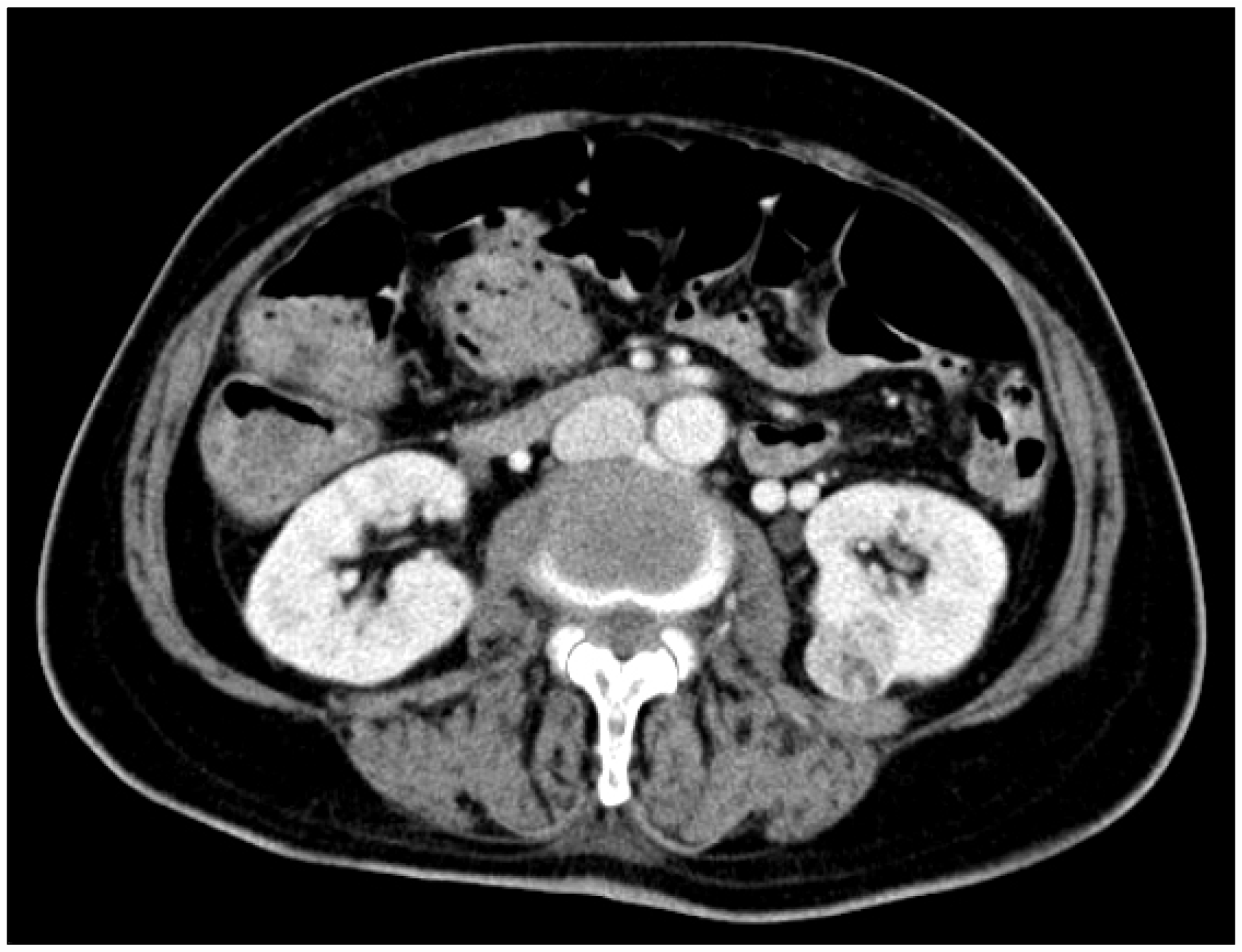
Fig. 2.
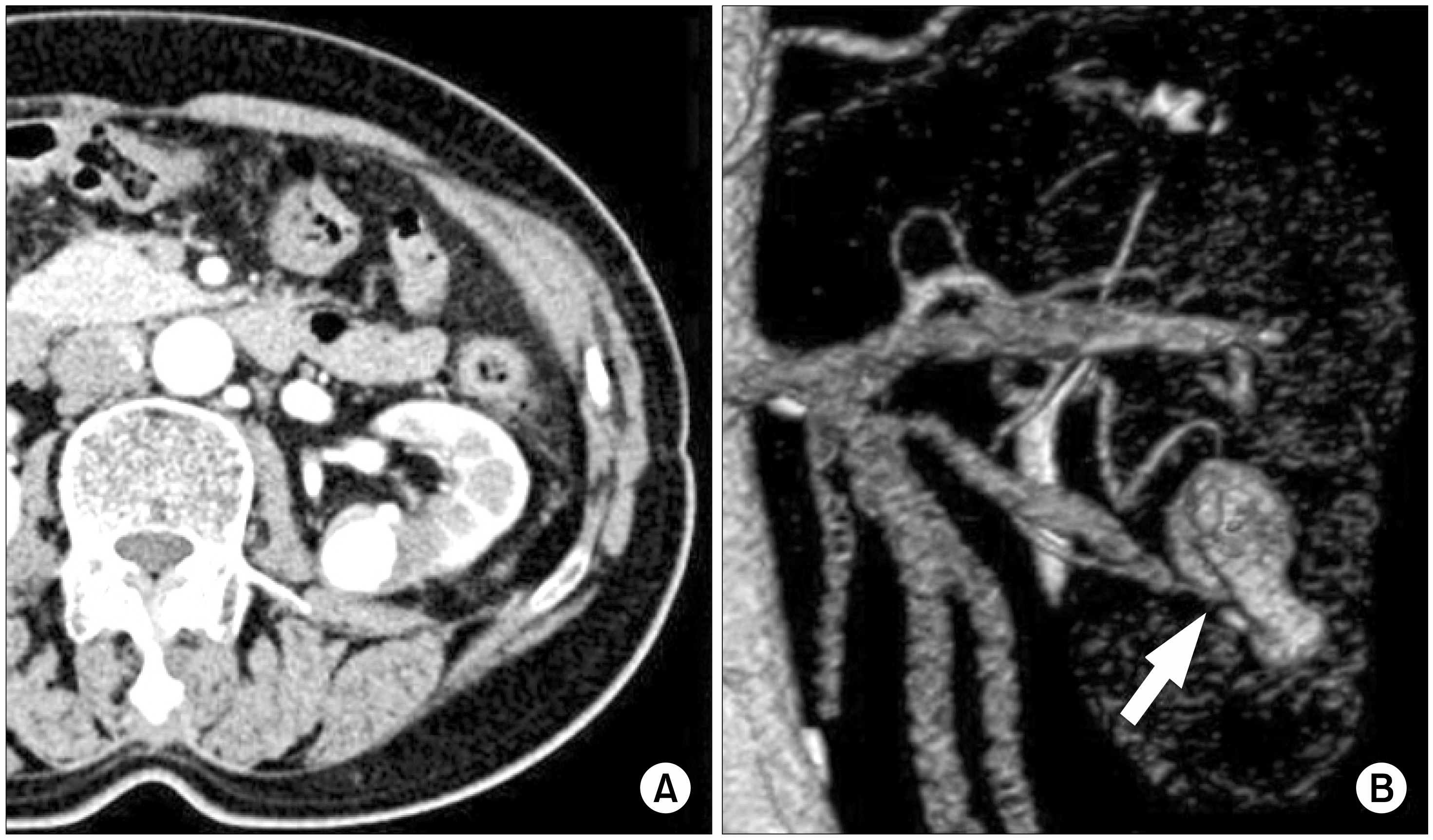
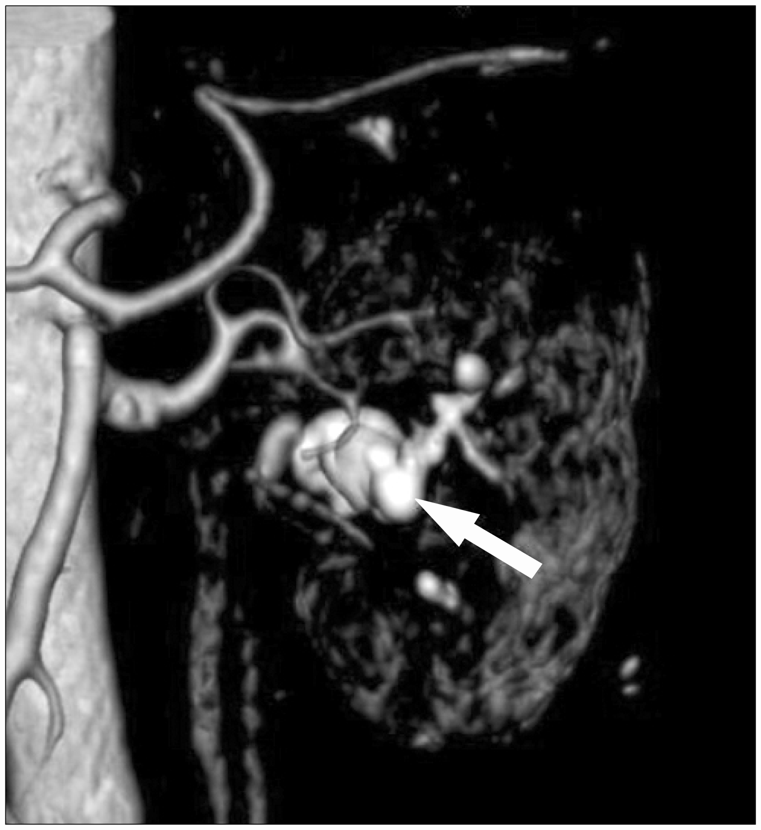
(A) Follow-up contrast CT scan of the kidney shows a large collection of contrast in the left lower renal pole, which was compatible with a pseudoaneurysm. (B) the 3-D volume rendered CT scan with intravenous contrast demonstrates a 3.5 cm sized saccular pseudoaneurysm (white arrow) arising from the lower segmental branch of the left renal artery.
 XML Download