REFERENCES
1. Hedrich CM, Fiebig B, Hauck FH, Sallmann S, Hahn G, Pfeiffer C, et al. Chilblain lupus erythematosus: a review of literature. Clin Rheumatol. 2008; 27:949–54.
2. Viguier M, Pinquier L, Cavelier-Balloy B, de la Salmonière P, Cordoliani F, Flageul B, et al. Clinical and histopathologic features and immunologic variables in patients with severe chilblains. A study of the relationship to lupus erythematosus. Medicine (Baltimore). 2001; 80:180–8.
3. Tüngler V, Silver RM, Walkenhorst H, Günther C, Lee-Kirsch MA. Inherited or de novo mutation affecting aspartate 18 of TREX1 results in either familial chilblain lupus or Aicardi-Goutières syndrome. Br J Dermatol. 2012; 167:212–4.

4. Abe J, Izawa K, Nishikomori R, Awaya T, Kawai T, Yasumi T, et al. Heterozygous TREX1 p. Asp18Asn mutation can cause variable neurological symptoms in a family with Aicardi-Goutieres syndrome/familial chilblain lupus. Rheumatology (Oxford). 2013; 52:406–8.
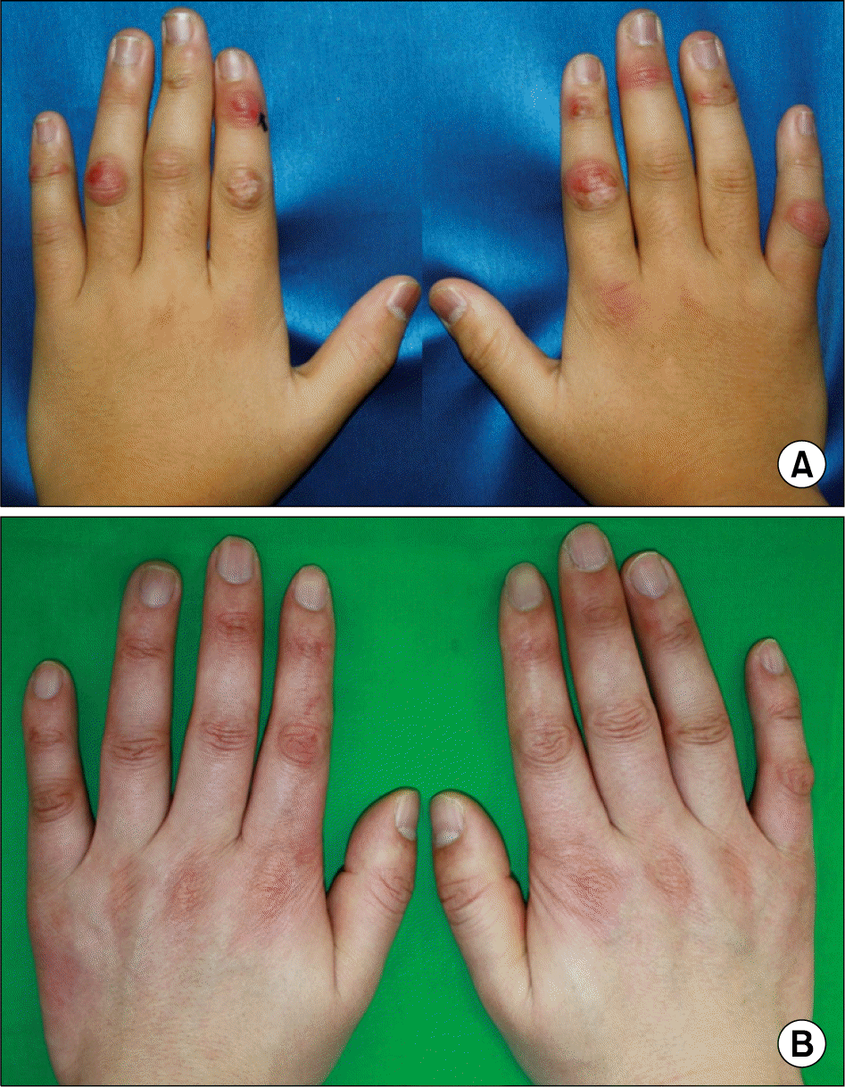
Figure 1.
(A) Multiple erythematosus scaly plaques were observed on dorsal aspects of patient's hands. (B) Similarily, his father showed erytheamtous skin lesions on dorsum of hands.

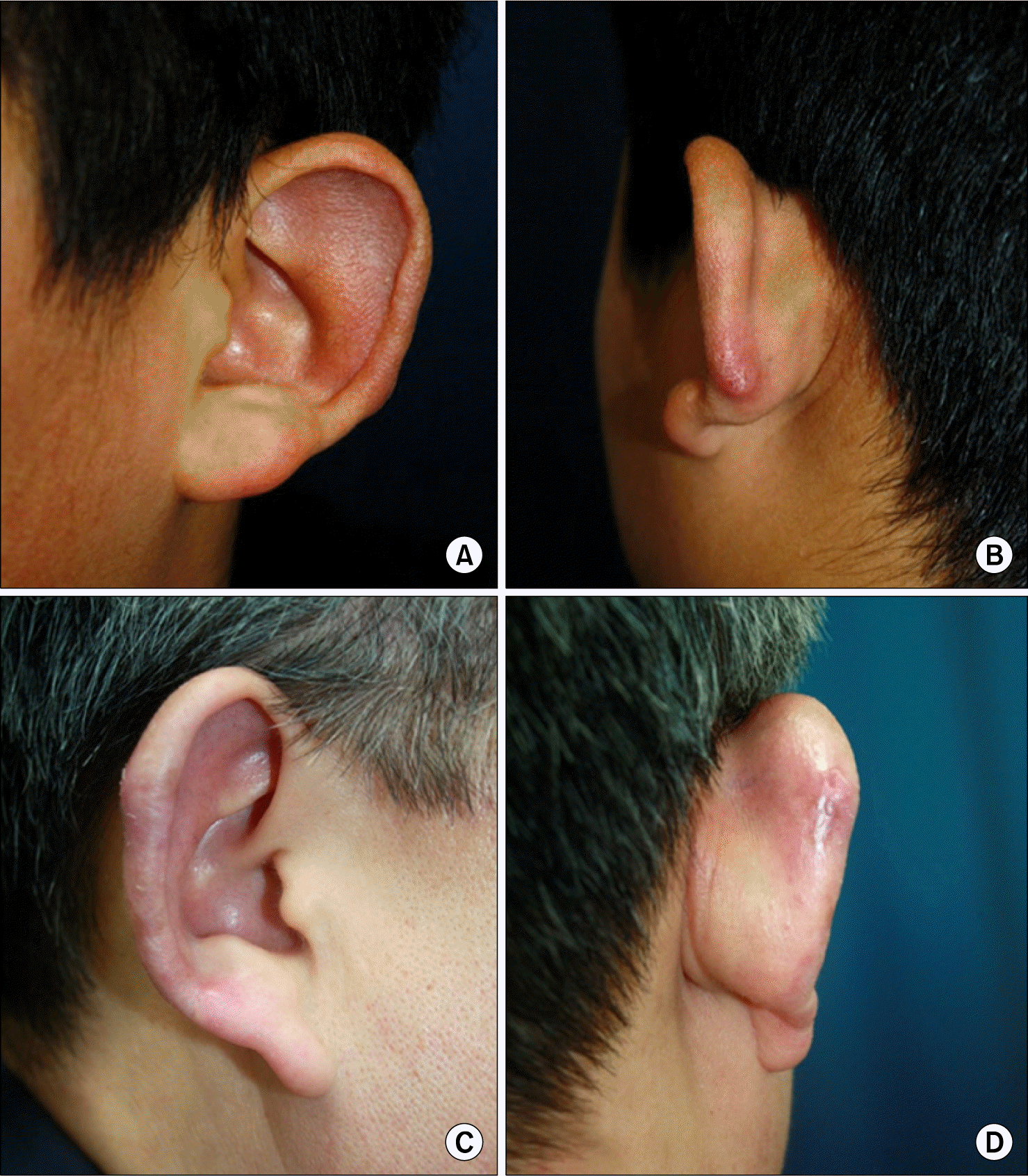
Figure 2.
Erythematous to pur-puric swollen scaly patches were noted on the ear of the patient (A, B) and his father (C, D).

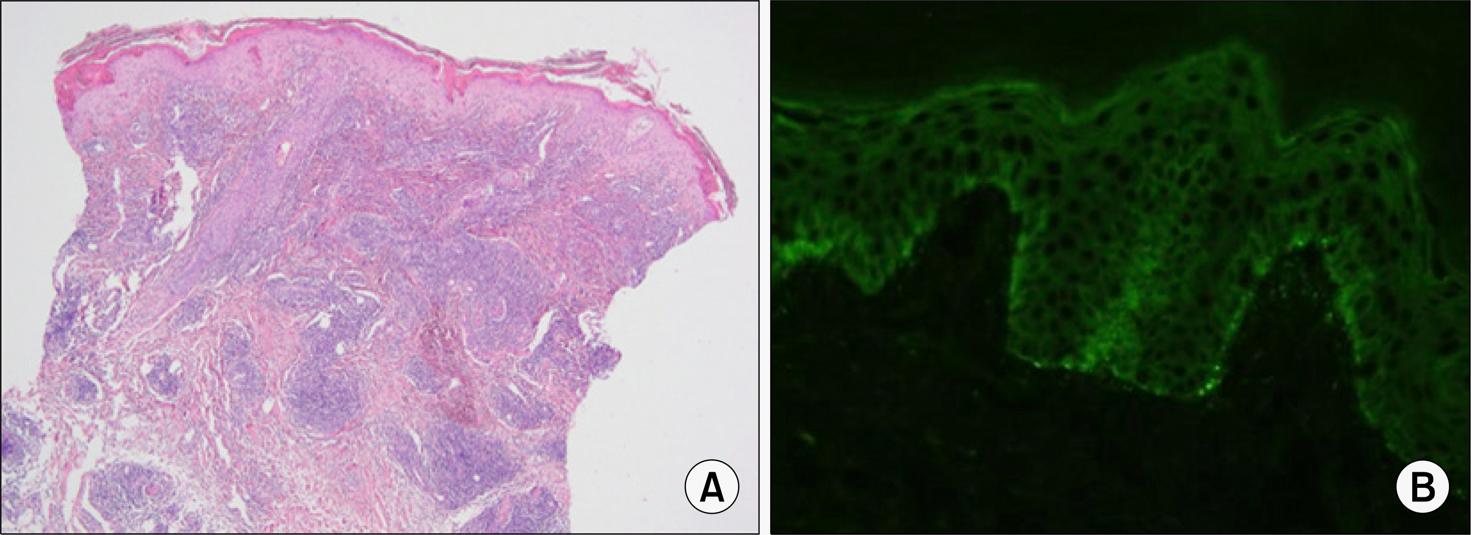
Figure 3.
(A) Histopathologic examination revealed interface dermatitis and prominent periap-pendigeal, perivascular and peri-neural lymphohistiocytic infiltrates throughout the entire dermis (H&E, ×40). (B) Lesional direct immunofluorescence results showed granular deposits of immunoglobulin M in the basement membrane zone (×200).


 PDF
PDF ePub
ePub Citation
 Print
Print


 XML Download