

 PDF
PDF ePub
ePub Citation
 Print
Print


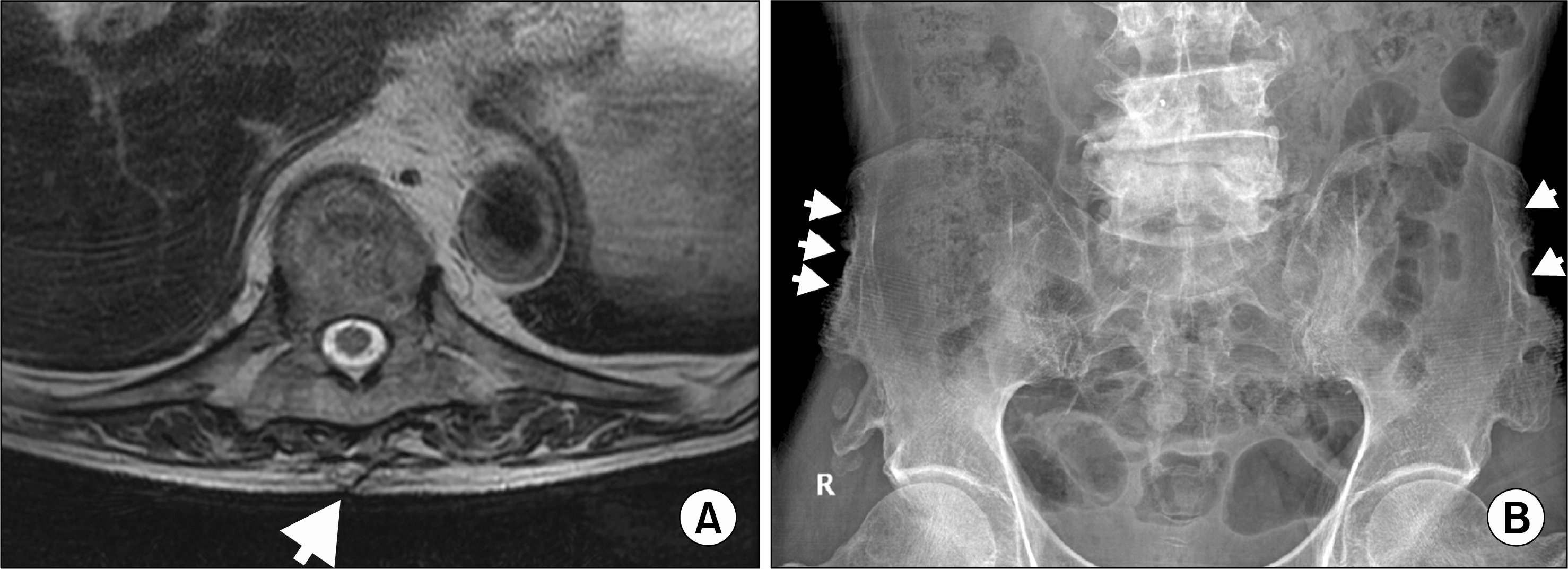
Figure 1.
(A) T-spine X-ray, lateral. (B) Spinemagnetic resonance imaging: Newly appeared compression fracture at T9 and segmental cord signal change at T9 level (white arrow head). Posterolateral fusion mass on T7-L1 that was misinterpreted as bony bridge were shown (white arrows). Additionally, bulging disc and facet arthrosis were shown at multiple level of spine.

 XML Download