

 PDF
PDF ePub
ePub Citation
 Print
Print


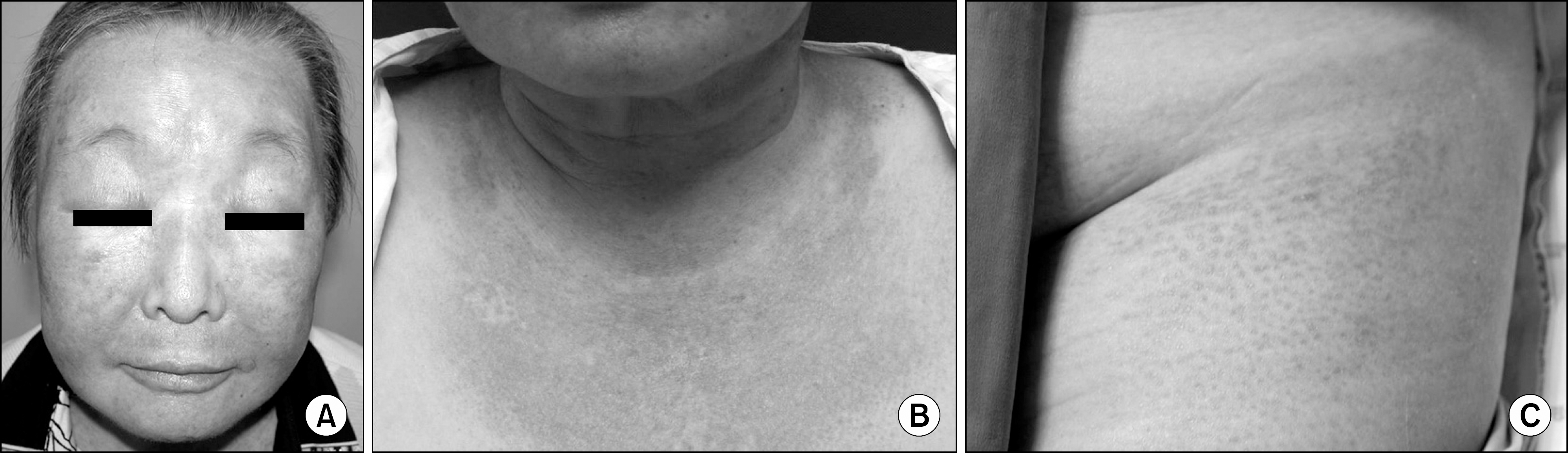
Abstract
Dermatomyositis (DM) is rare systemic inflammatory disease with typical skin manifestations and muscular involvement. Various skin lesions can accompany this disease, such as Gottron's sign, Heliotrope rash, mechanic's hands, V sign and shawl sign. Scleredema is a very rare skin manifestation in DM. We report a case of DM in a 63-year-old woman, who had scleredema on her thighs. A diagnosis of DM was established by clinical manifestation, elevated muscle enzyme levels, electromyogram measures, and muscle biopsy findings. She was successfully treated with the immunosuppressants methotrexate, cyclosporine, and steroids (low dose).
References
1. Ha YJ, Jung SY, Lee KH, Choi JJ, Lee KK, Lee SK, et al. A case of dermatomyositis showing vesicular lesion associated with ovarian cancer. J Korean Rheum Assoc. 2009; 16:291–5.

2. Chung JH, Seo PG. Clinical manifestations of dermatomyositis. Korean J Dermatol. 2002; 40:258–65.
3. Tanimoto K, Nakano K, Kano S, Mori S, Ueki H, Nishitani H, et al. Classification criteria for polymyositis and dermatomyositis. J Rheumatol. 1995; 22:668–74.
4. Jeong EC, Choi KH, Kim JH, Kim JH. A case of scleredema. Korean J Dermatol. 1985; 23:658–61.
5. Lee MW, Choi JH, Sung KJ, Moon KC, Koh JK. Scleredema: clinicopathological study. Korean J Dermatol. 2002; 40:119–23.
6. Cho B, Lee IJ. A case of scleredema. J Korean Pediatr Soc. 1993; 36:1770–3.
7. Mattheou-Vakali G, Ioannides D, Thomas T, Lazaridou E, Tsogas P, Minas A. Cyclosporine in scleredema. J Am Acad Dermatol. 1996; 35:990–1.
8. Seyger MM, van den Hoogen FH, de Mare S, van Haelst U, de Jong EM. A patient with a severe scleroedema diabeticorum, partially responding to low-dose methotrexate. Dermatology. 1999; 198:177–9.
9. Krasagakis K, Hettmannsperger U, Trautmann C, Tebbe B, Garbe C. Persistent scleredema of Buschke in a diabetic: improvement with high-dose penicillin. Br J Dermatol. 1996; 134:597–8.
10. Thumpimukvatana N, Wongpraparut C, Lim HW. Scleredema diabeticorum successfully treated with ultra-violet A1 phototherapy. J Dermatol. 2010; 37:1036–9.

 XML Download