



 PDF
 ePub
 Citation
 Print


INTRODUCTION
Asthma is a disease characterized by chronic airway inflammation, and the cause of inflammation is driven by gene and environment interactions. Although these interactions may be affected by many environmental factors, the most important environmental factors are indoor allergens of dust mite and outdoor air pollutants. Both risk factors may trigger the inflammation independently or synergistically. The successful identification of environmental allergens and avoidance of pollutants may be effective to prevent allergen marching and disease exacerbation.
HDM CAN BE IDENTIFIED IN THE HOUSEHOLD USING DIFFERENT METHODS
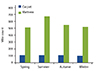
We used the floating method to identify the types of mites from houses of mite-allergic patients. The results showed that Dp accounted for 52.1% of the total number of mites and was found in every residence. Bt accounted for 44.3% of the total number of mites. We also demonstrated high prevalence of sensitization to Bt in Taiwan [4]. We used a series of recombinant allergenic components of Bt for specific IgE measurement. As well, Bt crude extracts were used for skin tests. We found that 44% of asthmatic patients were allergic to Bt. Furthermore, 43% of these Bt-allergic patients were also allergic to Blo t 5 [5]. A high prevalence of Bt sensitization was detected in asthmatic patients. However, 18% of these patients were caused by cross reactivity between Dp and Bt. In order to facilitate the analysis of IgE specific to Der p 2, the major allergen in Dp, we raised a panel of monoclonal antibodies (MoAbs) to Der p 2 antigens. We used MoAbs to purify major allergen Der p 2 from HDM extracts, and used the purified Der p 2 in skin tests and serology tests. The results showed that 90% of the patients' skin reacted to Der p 2 and their serum contained Der p 2-specific IgE. Thus, we demonstrated the incidence of Der p 2 hypersensitivity in asthmatic patients [6]. Furthermore, we can use these MoAbs to calculate the mite numbers in the dust by quantifying the concentration of Der p 2. In the most recent survey from April 2010 to March 2011 conducted by medical centers in central Taiwan, the numbers of mites in the carpets and mattresses were different [7]. There are a significantly higher number of mites in the mattresses than in the carpets. The number of mites also fluctuates with season, with the highest number of mites in the summer (Fig. 1). In the recent years, a novel technology has been discovered to identify environmental mite allergens. A highly sensitive nanostructured biosensor, including a three-dimensional (3D) sensing component with uniformly deposited gold nanoparticles, was used to detect Der p 2. The detection limit of the 3D gold nanoparticle-based biosensor was 1 pg/mL and the dynamic range was 5 µg/mL (Fig. 2). The novel nanostructured biosensor could be useful for fast detection of environmental allergens [8].
EXPOSURE TO HDM CAN CAUSE ALLERGIC RESPONSES IN THE AIRWAY OF BOTH HUMANS AND MICE
Dust mite can induce airway inflammation: In the allergic sensitization phase, dendritic cells (DCs) play an important role by acting as professional antigen presenting cells in the allergic airway inflammation [9, 10]. After the desquamation of epithelial cells and the disruption of tight junction caused by HDM, DCs can identify and process the antigens, and then present these antigens on the major histocompatibility complex class II molecules to naive T lymphocytes in local lymph nodes. This in turn induces cascades of the T helper 2 (Th2)-immune allergic inflammatory processes [10, 11, 12]. In the Dp-allergic subjects who were intranasally challenged with Dp extracts, both the immediate and late phases of allergic response were observed. HDM extracts could induce nasal symptoms by increasing nasal resistance [13]. When subjects were intranasally administrated with HDM extracts in animal study, a chronic airway inflammation was up-regulated [14]. Similar report showed that inhalation of HDM extracts significantly increased the recruitment of eosinophils and a substantial number of neutrophils, macrophages and lymphocytes [15]. With the characteristics of mixed eosinophilic and neutrophilic inflammation, the HDM mouse model becomes an interesting animal model that may closely reflect the situation of allergic asthma in humans [16].
IDENTIFICATION OF SM IN KITCHENS AND ITS CORRELATION WITH ALLERGIC DISEASES
SM has been reported to induce anaphylaxis through the consumptions of mite-contaminated food [17]. Based on its allergenic potential and abundance, the storage mite Tp is a major cause of allergic asthma and allergic rhinitis. Tp is found worldwide, mainly in contaminated food such as mushrooms, wheat flour, wheat germ, animal chow, and cereal (Table 1) [18]. Tp-induced allergy is mostly reported as occupational exposure among farmers, grain workers, bankers, and food industry workers, and is considered closely associated with respiratory diseases in these individuals [19].
EXPOSURE TO SM CAUSES AIRWAY INFLAMMATION IN MOUSE ANIMAL MODEL
Tp, a kind of SM, has been shown to induce allergic symptoms such as bronchial asthma, allergic rhinitis, conjunctivitis, and atopic dermatitis [20, 21]. In recent years, storage mites have been reported to be the key allergenic components of dust samples worldwide, thus highlighting their clinical significance [22].
In animal models, Balb/c mice were intraperitoneally sensitized with Tp and then intratracheally challenged with Tp. The airway inflammation was analyzed by airway hypersensitivity to methacholine and bronchial alveoli larvage immune responses to Tp were analyzed by immunoglobulin and cytokines measurements.
When Balb/c mice were challenged with different concentration of Tp (10 µg/µL and 40 µg/µL), there existed a dose-dependent increase of Tp-specific IgE and IgG1 in the sera. In addition, the inflammatory cell infiltration was up-regulated in the airway. Not only were the eosinophils and neutrophils increased, but the levels of Th2 cytokines, interleukin (IL) 4, IL-5, and IL-13 were also increased in the bronchial alveolar lavage fluid in the Tp-sensitized mice. This airway inflammation was associated with airway hypersensitivity to methacholine and the reverse of Th1/Th2 subpopulation in the blood. When the lung tissue was analyzed for the proinflammatory cytokine IL-6/IL-17F and transcription factor GATA-3, their levels were all elevated (Table 2) [23].
DIFFERENT AGE GROUPS OF SUBJECTS SENSITIZED BY DIFFERENT SPECIES OF HDM & SM
A previous study demonstrated that Dp is highly prevalent in Taiwan, where the climate is subtropical [24]. More than 80% of asthmatic patients are allergic to Dp as determined by skin tests and specific IgE measurements [25]. Dp and Tp have been demonstrated to be related to allergic diseases [24, 26]. Although Tp can cause allergic respiratory symptoms after occupational exposure in farms and grain elevator stores, a lot of attention is currently being paid to its allergenicity in nonoccupational environments [27].
There is a high prevalence of Tp sensitization in general adult populations in both Europe [27] and Asia [28]. In Spain, HDM-allergic patients also show a very high prevalence (73-83%) of storage mite sensitization [28]. Fifty-two percent of adult individuals over the age of 50 are sensitive to Tp, which is much more prevelent than Dp sensitization in the same age group (22%). Similar findings have been reported in Korea [29].
Subjects of different age groups were compared for their dust mite specific IgE in the sera. There were more subjects sensitized by Tp than Dp in the subjects over the age of 70 years. The association between age and sensitization to dust mites was different; there were more subjects below the age of 40 who were sensitized to Dp, and more subjects over the age of 70 that were sensitized to Tp (Table 3). The clinical relevance of mite sensitivity in elderly subjects with chronic obstructive pulmonary disease (COPD) was also analyzed. The results showed that, among the 63 elderly subjects, 50 (79.3%) had COPD. Among the 50 subjects with COPD, 38.0% (19/50) were Tp-sensitive subjects and 14.0% (7/50) were Dp-sensitive subjects [29]. All of the Dp-sensitive subjects were also Tp-sensitive. The results showed that the elderly subjects with COPD had a higher prevalence of sensitivity to Tp than to Dp, and the IL-8 level was significantly increased in allergic COPD patients (Table 4) [30].
DIFFERENT ALLERGENIC COMPONENTS OF MITES CAN ACTIVATE INNATE IMMUNE CELLS LEADING TO ALLERGIC INFLAMMATION
More than 21 allergenic components have been identified in the mites, most of which can initiate immune responses via pattern recognition receptor (Table 5).
Der p 1, a cysteine protease, can bind its specific IgE on the basophils and mast cells and can possess a very potent proteolytic activity on epithelium, resulting in a series of specific and nonspecific responses. Der p 1 can up-regulate the permeability of epithelial cells in vivo, most likely via degradation of tight junction proteins. In addition, a more specific response may be induced by these proteolytically active allergens. Their interaction with epithelial cells can induce the release of cytokines, such as IL-6 and IL-8, or induce fluid secretion from submucosal glands [31]. The mechanism underlying this response depends upon protease-activated receptors (PARs). PAR 2 are activated by proteolytic cleavage, which can be mediated by Der p 1 [32, 33]. Thus, Der p 1 may serve as the prototype of proteolytically active allergens.
Der p 2 and its specific IgE in the sera are highly correlated with allergic hypersensitivity in patients of asthma, atopic dermatitis and allergic rhinitis. An estimate of 79.2% of patients with asthma, wheezing and/or rhinitis has IgE antibodies to Der p 2 [34]. Recently, the Der p 2 allergen was found to show structural homology with MD-2, suggesting that Der p 2 tends to cause targeted immune responses because of its autoadjuvant properties. The structure of Der p 2 provides a useful tool in the design of recombinant immunotherapeutics for the group-2 allergens [35]. Der p 2 has been demonstrated to be capable of triggering human B cell activation and Toll-like receptor 4 (TLR4) induction. Der p 2 markedly induced the expressions of several key cytokines, including IL-1β, CXCL10, IL-8, and tumor necrosis factor (TNF)-α in B cells. Der p 2 could also enhance nuclear factor (NF)-kB activity, indicating that Der p 2 may exert its influence through NF-kB activation to induce the production of proinflammatory cytokines. Moreover, Der p 2 specifically up-regulated mitogen-activated protein kinase phosphatase-1 expression and activity in human B cells, which in turn resulted in p38/mitogen-activated protein kinase dephosphorylation, triggering TLR4 induction [36].
Der p 3 can induce cytokine release from human pulmonary epithelial cells. Because Der p 3 is a serine protease, it is likely to interact with the PAR 2 expressed on pulmonary epithelial cells [37]. Like trypsin, a known activator of PAR 2, Der p 3 cleaved the receptor at a specific site that exposes the tethered ligand, indicating that they do have the potential for activating PAR 2. In addition, Der p 3 can induce the release of the proinflammatory cytokines granulocyte-macrophage colony-stimulating factor (GM-CSF) and eotaxin from human pulmonary epithelial cells. The production of both GM-CSF and eotaxin is increased in bronchial epithelial cells from asthmatic patients, and these cytokines play a major role in the ongoing inflammatory and structural alterations of asthma [38, 39, 40, 41, 42]. Eotaxin and GM-CSF promote accumulation local activation of eosinophils in the bronchial mucosa of asthmatic patients [40, 41, 42, 43]. We also demonstrate group 3 allergen of Tp (Tyr p 3) had proteolytic activity and specific IgE in the sera from patients with airway hypersensitivity [26].
HDM ALLERGY: DISEASE PREVENTION
Interventions to reduce allergen sensitization and to improve patients' symptoms and quality of life have obtained great achievements. However, prevention strategies for treatment of allergic diseases remain unsuccessful. Recent evidence from clinical studies has highlighted different nutritional interventions, such as dietary polyphones vitamin D, which can impact both allergic sensitization and alleviated symptoms [44, 45]. The hygiene hypothesis suggested that sensitization may have affected the intestinal microflora composition and the immune system at an early age [46]. Distinct patterns of gut microbial composition were reported in infants with and without allergy development [47]. Supplement of probiotics is the best modality for the prevention of HDM allergy.
According to Cochrane review by Osborn and Sinn [48] in 2007, probiotics in infants for prevention of allergic diseases and food hypersensitivity was not supported by sufficient evidence. Many further investigations have been done since then. In 2013, Pyar et al. [49] indicated many diseased conditions have been observed to be due to in vivo microbial imbalance and the consequent conditions could be remedied by the application of appropriate probiotics. Probiotics can protect infants against life-threatening diseases by improving their bowel colonization with beneficial flora through the stimulation of the immune system. In the recent study, Vong et al. [50] demonstrated that probiotic Lactobacillus rhamnosus strain GG inhibits both phorbol 12-myristate 13-acetate- and Staphylococcus aureus-induced formation of neutrophil extracellular traps, which can prevent pathogens from damaging surrounding cells and can be used to treat chronic inflammation bowel disease. Moreover, probiotic L. rhamnosus strain GG could dampen reactive oxygen species production and modulate innate immunity response of the gastrointestinal tract. Therefore, whether probiotics could prevent allergic disease was reviewed.
ALLERGIC SENSITIZATION CAN BE PREVENTED BY INCREASING T REG CELLS AND SECRETORY IGA
The pathogenesis of allergic diseases is considered to be Th2-mediated inflammation. Medical scientists have hypothesized that allergic diseases might result from functional or quantitative deficiency of Treg cells that control the Th2 immune response [51]. Wu et al. [52] reported that there were fewer Treg cells in allergic patients in comparison with nonallergic subjects. These cells could be up-regulated by using recombinant Der f 2 peptide with fungal immunomodulatory peptide fve (FIP).
It has been reported that allergen specific IgA could prevent the increased airway responsiveness and lung eosinophilia in allergen sensitized mice [53]. Intranasal immunotherapy is more effective than intradermal immunotherapy for the induction of airway allergen tolerance in Th2-sensitized mice [54]. Our study also showed that the production of salivary IgA and suppression of Dp-induced airway inflammation were observed after local nasal immunotherapy with Dp2 in conjunction with FIP. We used local nasal immunotherapy with Der p 2 in conjunction with FIP [55]. From the above studies, increased IgA levels could down-regulate airway hypersensitivity, Th2-associated cytokine release and IgE production.
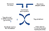
Administration of probiotics could stimulate the immune system and reduce inflammation on gastrointestinal diseases by increasing Treg cytokines [56]. Chiua et al. [57] reported that cytokines of Treg cells, IL-10 and TGF-β, could be up-regulated by probiotics. Therefore, it is conceivable that probiotics administration could be beneficial for allergic diseases (Fig. 3).
EFFECTS OF PROBIOTICS ON IGA PRODUCTION
Infants with allergy development have different patterns of gut microbial composition when compare to nonallergic subjects. The presence of Clostridium difficile indicated a high risk of developing eczema and allergic sensitization [58]. Supplemental administration can promote in situ inhibition of pathogens through IgA secretion of the host to reduce allergic allergen sensitization and symptoms. The level of circulating IgA was increased after probiotic intervention. Dolle et al. [59] reported that EcN, serotype O6:K5:H1 and a gram-negative bacteria strain, could up-regulate serum IgA levels in grass allergic patients. Similar report showed that consumption of probiotic products containing Escherichia coli strains Lactobacillus gasseri CECT5714 and Lactobacillus coryniformis CECT5711 could induce beneficial effects on immunological parameters in allergic children, causing a reduction in plasma level of IgE and an increase in the level of IgA. The probiotic dairy products increased IgA and improved the general health status of children [60].
On the other hand, probiotics failed to increase secretory IgA levels in the breast milk of these subjects during pregnancy. As Boyle et al. [61] revealed in their experiment, 250 pregnant women carrying infants at high risk of allergic diseases were prenatally treated with the probiotic L. rhamnosus GG (LGG) for the thirty-six week gestation until delivery. The results showed that prenatal probiotic treatment was not associated with a reduced risk of eczema or IgE-associated eczema. Furthermore, prenatal probiotic treatment was not associated with any change in cord blood immune markers, but was associated with decreased soluble CD14 and IgA levels in breast milk. The study could not provide evidence that the probiotic had beneficial effects on IgE-associated eczema. This result might be due to the low IgA level in the breast milk [62].
EFFECTS OF PROBIOTICS ON TREG CELLS
Yoshida et al. [63] used Balb/c mice which were administered Lactobacillus plantarum NRIC0380 orally, and found that the increased proliferation of Treg cells was obtained from the mice that had been fed NRIC0380. The Treg cells obtained from the mice exposed to NRIC0380 significantly inhibited the IgE production and active cutaneous anaphylaxis reaction when transferred into another mouse that was subsequently immunized with the crude bovine β-lactoglobulin. Furthermore, the Treg cells also significantly suppressed the passive cutaneous anaphylaxis reaction when they were cotransferred with the IgE antibody into another mouse. In ovalbumin (OVA) immunized mice modal, L. rhamnosus (Lcr35) treatment of Ha-Jung Kim et al. [64]. led to an increase in the number of CD4+ CD25+ Foxp3+ Treg cells in the mesenteric lymph nodes of allergic march in OVA sensitized mice. Similar finding of Lcr35 treatment was observed in the report of Jang et al. [65]. In their experiment, Balb/c mice were administered Lcr35 a week before first sensitization, and airway hyperresponsiveness, total IgE production, pulmonary eosinophilic inflammation, and splenic lymphocyte proliferation were suppressed after Lcr35 treatment. Th1 (interferon [IFN]-γ) and Th2 (IL-4, IL-5, and IL-13) cytokines in the serum were also suppressed, and the percentage of Treg cells in the spleen was significantly increased in the Lcr35 treated group. Anti-CD25 mAb administration abolished the protective effects of Lcr35, indicating that CD4+ CD25+ Foxp3+ Treg cells are essential in mediating the activity of Lcr35. In the above animal model of allergic diseases, probiotics could increase Treg cells and down-regulate Th2 cell cytokine and IgE production.
BENEFIT OF PROBIOTIC ON THE ALLERGIC SYMPTOMS REMAINS INCONCLUSIVE
We reviewed current publications in the recent four years. Studies of probiotics on allergic respiratory symptoms are still inconclusive. A few studies conducted in children and adults with allergic rhinitis had encouraging results showing that allergic rhinitis symptoms could be reduced. Effective reports were conducted in several double-blind placebo controlled studies, which demonstrated reduced allergic rhinitis symptoms and down-regulated Th2 cells activation following treatment with Lactobacillus paracasei NCC2461, Lactobacillus salivarius PM-A0006, Lactobacillus johnsonii EM1 and L. paracasei ST11 in children or adolescents with allergic rhinitis (Table 6) [66, 67, 68, 69]. However, some studies conducted in adults were not effective in reducing the symptoms. Noneffective reports were conducted in the studies, which included the failure to relieve allergic symptoms in children or adults given L. paracasei, Bifidobacterium longum (BL999), L. rhamnosus (LPR), Lactobacillus casei Shirota, L. paracasei ssp paracasei F19 (Table 7) [70, 71, 72, 73]. These different outcomes could be due to different strains of probiotics used. To assure the effect of probiotics on allergic inflammation, the optimal dosage, probiotic strains, duration of intervention and patients' subpopulations were important.
DIFFERENT STRAINS OF PROBIOTICS HAVE DIFFERENT FUNCTIONS
Since Human breast milk contained a wide spectrum of indigestible nutrients that are not utilized by the infants but exert biomarker functions on establishment of indigenous microbiota, they might serve as natural prebiotics. The best examples are human milk oligosaccharides, which serve as growth stimulants for some colonic bacteria in breast milk as well as in breast-fed infants' gastro intestinal tract. Bifidobacteria are the predominant components of the intestinal flora in breast-fed infants. These bacteria groups play a vital role in maintaining the gut health and protecting the infants from pathogen infections, and contribute to maturation of the systemic immunity.
A total of 18 strains of bacteria strain were identified from human breast milk. We found that Bifidobacterium adolescentis DB-2458 and Bifidobacterium longum subsp. infantis GB-1496 exhibited a strong immunomodulatory ability to increase Th1-associoated cytokine induction and to lower Th2-associated cytokine production. Besides these two strains, B. longum HB-762 exerted the most excellent anti-inflammatory capacities according to high levels of IL-10 and TGF-β cytokine inductions [57]. Similar finding was found in L. casei MYL01, which induced the production of IL-10 from HepG2 cells [74].
Eleven Bifidobacterium strains isolated from the feces of newborn babies were screened for their ability to induce cytokine production of human peripheral blood mononuclear cells. Among the eleven viable strains, L. gasseri AI-88 was the strongest inducer of IFN-γ and IL-12p70 production. However, after heat treatments, its stimulatory ability was attenuated. Heat-killed Enterococcus faecalis YM-73 and L. salivarius AP-32 strains showed an enhanced stimulation of IFN-γ and IL-12p70 secretion and a coincidental decrease in IL-13 production. The adhesion of lactic acid bacteria to Caco-2 cells decreased with the increase in temperature. However, heat exposure did not influence immunomodulatory activity. As temperature rose, roughness and unevenness of bacterial cell surfaces increased significantly. The results indicated that heat-killed E. faecalis YM-73 and L. salivarius AP-32 possessed immunomodulatory ability by increasing Th1-associated cytokines and reducing Th2-associated cytokines, switching the immune response from a Th2 toward a Th1 response [75].
In clinical studies, four strains of probiotics, B. adolescentis DB-2458, B. longum subsp. Infantis GB-1496, B. longum HB-762, and Lactobacillus reuteri GL-44, were used to evaluate their effects on the peripheral mononuclear cells derived from eight HDM allergic patients. The results demonstrated that Th2 associated cytokines, IL-5 and IL-13, and proinflammatory cytokines, IL-6 and TNF-α, were both decreased. In the presence of Dp crude extract, probiotics-DB-2458, GB-1496, and HB-762-could improve the balance of Th1/Th2 and reduce the production of inflammatory cytokines from allergic subjects.
In the above studies, probiotics isolated from breast milk and infant feces could up-regulate Th1 associated cytokine and reduce Th2 associated cytokine to prevent allergy development. Both freshly isolated and heat-killed bacilli were similarly effective in the in vitro study.
CONCLUSION
Dust mite allergy affects approximately 40% of the industrial global population. It could be found in the living and working environments. There are many types of dust mite species and prevalence of different mite allergy varies with patients' age groups. Major mechanism of allergic response is Th2 cytokine production, which would induce the production of IgE antibodies by B cells. When the subjects are exposed to allergens again, allergic reactions would be induced. Since using drugs such as steroid, antihistamine, etc. are not successful. Therefore, prevention of allergic development is more important than treatment. Probiotics may provide an alternative modality to prevent allergy developments and to achieve beneficial effects by increasing the number of Treg cells and by enhancing IgA production from B cells. Although the effect of probiotics is still inconclusive on allergic symptoms, in order to achieve successful interventions, choosing correct therapeutic doses and strains of microbiota could be critical to achieve optimal therapeutic effects.









 XML Download