Abstract
Objective:
Patients undergoing intracranial operations often suffer from post-operative epidural hematoma (EDH). The incidence and risk factors for with the occurrence of EDH after intracranial operations are not well described previously. The objective of this study was to identify the risk factors and the incidence of post-operative EDH adjacent and regional to the craniotomy.
Methods:
This was a retrospective study of 23 (2.4%) patients, between January 2005 and December 2011, who underwent epidural hematoma evacuation after primary intracranial during this period, 941 intracranial operations were performed. The control group (46 patients) and hematoma group (23 patients) were categorized on the basis of having undergone the same pre-operative diagnosis and treatment within 3 months of their operations. The ages of the hematoma and control group were individually matched to similar ages within 10 years of each other to minimize bias of age.
Results:
Univariate analysis showed that the significant pre-operative and intra-operative factors associated with postoperative EDH were a pre-operative Glasgow Coma Scale (GCS) scored <8 (crude odds ratio 8.295), prothrombin ratio >1.0 (p=0.014), prothrombin time (PT) >11.3 sec (p=0.008), intra-operative blood loss >650 mL (p=0.003) and craniotomy size >7,420 mm2 (p=0.023). In multivariate analysis, intra-operative blood loss exceeding 650 mL (median of total patients) placed a patient at significantly increased risk for post-operative EDH.
REFERENCES
1). Altschuler E., Moosa H., Selker RG., Vertosick FT Jr. The risk and efficacy of anticoagulant therapy in the treatment of thromboembolic complications in patients with primary malignant brain tumors. Neurosurgery. 27:74–76. discussion 77. 1990.

2). Awad JN., Kebaish KM., Donigan J., Cohen DB., Kostuik JP. Analysis of the risk factors for the development of post-operative spinal epidural haematoma. J Bone Joint Surg Br. 87:1248–1252. 2005.

3). Ban SP., Son YJ., Yang HJ., Chung YS., Lee SH., Han DH. Analysis of complications following decompressive craniectomy for traumatic brain injury. J Korean Neurosurg Soc. 48:244–250. 2010.

4). Fukamachi A., Koizumi H., Nagaseki Y., Nukui H. Postoperative extradural hematomas: computed tomographic survey of 1105 intracranial operations. Neurosurgery. 19:589–593. 1986.

5). Garber ST., Sivakumar W., Schmidt RH. Neurosurgical complications of direct thrombin inhibitors—catastrophic hemorrhage after mild traumatic brain injury in a patient receiving dabigatran. J Neurosurg. 116:1093–1096. 2012.

6). Gentleman D., Johnston RA. Postoperative extradural hematoma associated with induced hypertension. Neurosurgery. 17:105–106. 1985.

7). Gerlach R., Raabe A., Zimmermann M., Siegemund A., Seifert V. Factor XIII deficiency and postoperative hemorrhage after neurosurgical procedures. Surg Neurol. 54:260–264. discussion 264-265. 2000.

8). Guangming Z., Huancong Z., Wenjing Z., Guoqiang C., Xiaosong W. Should epidural drain be recommended after supratentorial craniotomy for epileptic patients? Surg Neurol. 72:138–141. discussion 141. 2009.

9). Haft H., Liss H., Mount LA. Massive epidural hemorrhage as a complication of ventricular drainage. J Neurosurg. 17:49–54. 1960.

11). Hussain SA., Selway R., Harding C., Polkey CE. The urgent postoperative CT scan: a critical appraisal of its impact. Br J Neurosurg. 15:116–118. 2001.
12). Institute SAS. The sas system for windows, version 9.2. Cary, NC: SAS Institute. 1996.
13). Jeon JS., Chang IB., Cho BM., Lee HK., Hong SK., Oh SM. Immediate postoperative epidural hematomas adjacent to the craniotomy site. J Korean Neurosurg Soc. 39:335–339. 2006.
14). Kalfas IH., Little JR. Postoperative hemorrhage: a survey of 4992 intracranial procedures. Neurosurgery. 23:343–347. 1988.

15). Le Roux AA., Nadvi SS. Acute extradural haematoma in the elderly. Br J Neurosurg. 21:16–20. 2007.

16). Manian FA., Meyer PL., Setzer J., Senkel D. Surgical site infections associated with methicillin-resistant Staphylococcus aureus: do postoperative factors play a role? Clin Infect Dis. 36:863–868. 2003.
17). Palmer JD., Sparrow OC., Iannotti F. Postoperative hematoma: a 5-year survey and identification of avoidable risk factors. Neurosurgery. 35:1061–1064. discussion 1064-1065. 1994.
18). Park JE., Kim SH., Yoon SH., Cho KG., Kim SH. Risk factors predicting unfavorable neurological outcome during the early period after traumatic brain injury. J Korean Neurosurg Soc. 45:90–95. 2009.

19). Rapanà A., Lamaida E., Pizza V. Multiple postoperative intracerebral haematomas remote from the site of craniotomy. Br J Neurosurg. 12:364–368. 1998.
20). Sinar EJ., Lindsay KW. Distant extradural haematoma complicating removal of frontal tumours. J Neurol Neurosurg Psychiatry. 49:442–444. 1986.

21). Soff GA. A new generation of oral direct anticoagulants. Arterioscler Thromb Vasc Biol. 32:569–574. 2012.

22). Sokolowski MJ., Garvey TA., Perl J 2nd., Sokolowski MS., Cho W., Mehbod AA, et al. Prospective study of postoperative lumbar epidural hematoma: incidence and risk factors. Spine (Phila Pa 1976). 33:108–113. 2008.
23). Thiagarajah S. Postoperative care of neurosurgical patients. Int Anesthesiol Clin. 21:139–156. 1983.

24). Troupp H. Extradural hematoma during craniotomy. Report of five cases. J Neurosurg. 40:783–785. 1974.
FIGURE 1.
Results of computed tomography (CT) examinations. A: CT scan showing intracranial hematoma at right basal ganglia with midline shifting. B: Post-operative immediate brain CT scan showed almost completely removed intracerebral hemorrhage, and also showed large amount of epidural hematoma (EDH) with aggravated mass effect. C: After reoperation was performed to remove the EDH beneath the previous craniectomy site, brain CT scan showed no residual epidural hematoma. A marginal amount of intracerebral hemorrhage remained.

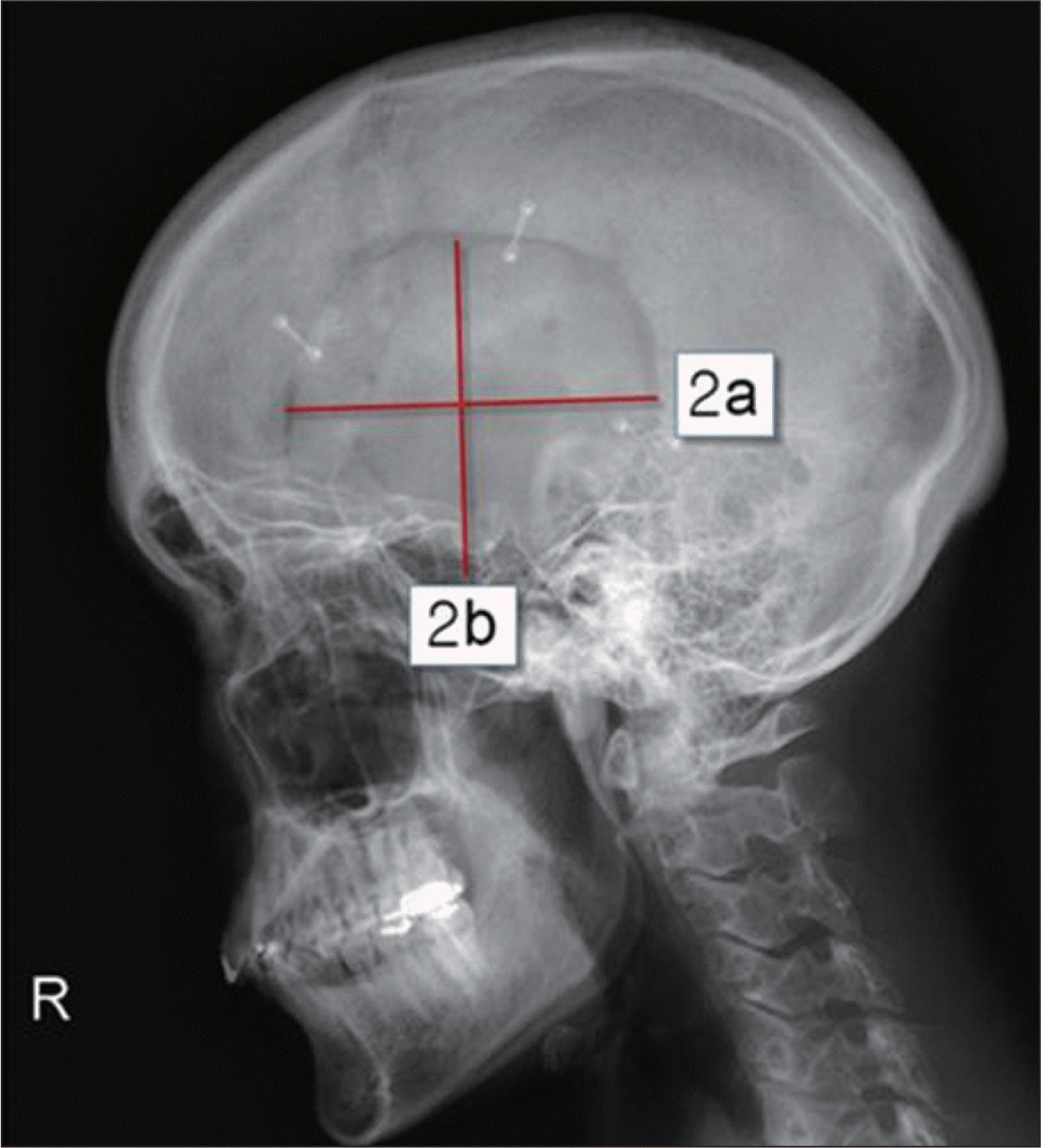
FIGURE 2.
Data concerning craniotomy extent. Craniotomy extent was measured from plain lateral skull X-ray that is, from maximal perpendicular diameter (radius a and b) of the craniotomy. The resulting size of craniotomy was then approximated using the formular for a circle (size=π×ab).

TABLE 1.
Clinical characteristics of the control and hematoma groups
TABLE 2.
Univariate conditional logistic regression analysis (pre-operative patient details)
TABLE 3.
Univariate conditional logistic regression analysis (pre-operative laboratory data)
TABLE 4.
Univariate conditional logistic regression analysis (intra-operative data)
TABLE 5.
Multivariate conditional logistic regression analysis
TABLE 6.
Correlation analysis of the parameters associated with coagulopathy and intraoperative blood loss exceeding 650 mL
| Estimated blood loss above 650 mL (n=35) | ||
|---|---|---|
| Pearson's r | p-value | |
| Prothrombinpercent (%) | 0.140 | 0.424 |
| Prothrombinratio (INR) | 0.061 | 0.727 |
| aPTT | -0.067 | 0.701 |
| Platelet count (×109/L) | 0.045 | 0.798 |


 PDF
PDF ePub
ePub Citation
 Print
Print


 XML Download