

 PDF
PDF ePub
ePub Citation
 Print
Print


I. INTRODUCTION
Complete obturation of the root canal system is the one of the keys of success in endodontic treatment, because the primary aim of root canal treatment is the elimination and future exclusion of all microorganisms from the root canal system. A root canal sealer, or cement, is used in combination with gutta-percha that is the most widely used and accepted obturating material. The sealer has a primary role in sealing the canal by obliterating the irregularities between the canal wall and the core material. Although many different filling techniques and materials have been used in order to obtain a adequate seal, a complete seal of the entire root canal is impossible1) .
The smear layer is a layer of sludge material formed over the surface of dentinal wall whenever dentin was cut. This layer may contain inorganic and organic materials, such as remnants of odontoblastic processes, necrotic pulp tissue and microorganism. Therefore it acts as an intermediate physical barrier and may interfere with adaptation of obturation materials to prepared root canal surfaces and penetration of sealers into the irregularities of root canal system and dentinal tubules2).
Several studies3-5) reveal that the removal of smear layer with EDTA improved the quality of apical sealing when AH26 sealer (Dentsply, Konstanz, Germany) was used.
The dental adhesive system is proposed to achieve a micromechanical bond to dentin, known as the hybrid layer, producing high bond strengths6). The resin tags of dentin bonding agent penetrate into the dentinal tubules. This hybrid layer can reduce the microleakage not only in restorative dentistry but also in root canal treatment. However, difficult methods of delivery into the root canal system and the inability to retreat the canal filled with resin alone if necessary are the main reasons for not using a dentin adhesive as a root canal filling material.
Mannocci and Ferrari7) tested a combination of the dentin bonding agent and epoxy-resin based root canal sealer to fill the canal system with lateral condensation technique. The root canal dentin showed the penetration of resin into the dentinal tubules and the apical sealing ability was better when compared with AH26 and gutta-percha. Leonard et al.8) also evaluated the characteristic hybrid layer of root canal dentin by scanning electron microscope.
The purpose of this study was to compare the sealing ability of epoxy resin-based sealer used in conjunction with two different dental adhesives and epoxy resin-based sealer alone with or without smear layer using an anaerobic bacterial leakage model.
II. MATERIALS AND METHODS
Sample Preparation
Fifty-two single-rooted human teeth with a single root canal, extracted for periodontal and orthodontic reasons, were used in this study. Superficial debris was removed from the roots with an ultrasonic scaler and the teeth were stored in physiologic saline until ready for use. The crowns were removed at the cementoenamel junction using a water-cooled tapered diamond bur. The working length was determined by passing a 15 K-type file into the canal until the tip was just visible at the apical foramen and then subtracting 0.5mm from this length.
The canals were cleaned and shaped with .04 and .06 taper Profile (Dentsply-Maillefer, Ballaigues, Swiss) using a crowndown pressureless technique. The apical portion of canal was enlarged to minimum ISO size #30 and maximum ISO size #55 file, depending on the size of original canal size. The apical patency was checked by passing a size #15 file through the apex.
Copious irrigation with 3.5% sodium hypochlorite was used throughout the cleaning and shaping of the canals using a syringe and 27 gauge endodontic needle. The canals were then flushed with 10ml of deionized water and dried with paper points. The prepared roots were randomly divided into four experimental groups of 12 teeth each. Four teeth were served as negative and positive controls and were prepared in the same manner as the experimental groups and received no filling (Group 5 and 6). Two teeth were used as negative controls (Group 6) and these teeth were completely coated with two coats of nail varnish.
The teeth in Group 1 were filled with gutta-perch and AH26 after All Bond 2 dental adhesive (Bisco, Itasca, IL, USA) application by the Continuous Wave of Condensation Technique. The canal walls were etched with 37% orthophosphric acid for 15 seconds, rinsed with 10ml-deionized water and dried with paper points. AH26 was then prepared according to the manufacture's directions. All Bond 2 primer was applied inside the canals. All Bond 2 Dentin Enamel Bonding Resin and Pre-Bond Resin was mixed and applied into the canal on a soaked paper point. The master gutta-percha point was coated with AH26 sealer and seated in the canal to the full working length. The root canal down-packed with System B (Analytic Endodontics, Orange, CA, USA) and backfilled with Obtura II (Obtura Spartan, Fenton, MO, USA) by the continuous wave of condensation technique. The appropriately tapered System-B Heat Plugger is preheated 200℃.
The teeth in Group 2 were filled with gutta-percha and AH26 after Prime & Bond NT Dual Cure Dental Adhesive System (Dentsply, Konstanz, Germany) application. The canal dentin was etched with Caulk 34% Tooth Conditioner Gel for 15 seconds. This was immediately rinsed for 10 seconds with 10ml deionized water and dried with paper points. Prime & Bond NT Dual Cure Dental Adhesive System was mixed according to the manufacturer's instructions and applied in the canal. Root canal filling procedure was then performed as described for the teeth in Group 1.
The smear layer of the teeth in Group 3 was removed by slowly injecting 10ml of 17% EDTA solution into the canal over a 5-minute-period. This was then followed by flushing the canal with 10ml of 3.5% NaOCl. No adhesive system was used in this group. Except this, the canal filling procedure was performed as described for the teeth in Group 1 and 2.
The root canals of Group 4 were filled with gutta-percha and AH26. There was no treatment of EDTA. The smear layer was still remaining. The root canals were filled by same procedure of Group 1 and 2.
The coronal ends of gutta-percha were cut off until a uniform filling length of 7mm was remained for all roots. After obturation, all roots were stored at 100% humidity and 37℃ incubator for 3 days to allow full setting of the sealer and adhesives.
Bacterial Leakage Model Preparation
A dual chamber anaerobic bacterial leakage model was assembled using a 5ml irrigation syringe and tooth as the upper chamber and a 20 ml scintillation vial (Samwoo Scientific Co., Seoul, Korea) as the lower chamber. The tooth was attached with cyanoacrylate cement and silicon glue to the tip of the syringe and the joint was sealed with two coats of nail varnish. The syringe was secured via a hole drilled through the cap of a 20 ml scintillation vial.
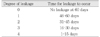
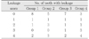
The apparatus was sterilized with ethylene oxide gas. The vials were placed in the anaerobic chamber (Coy Laboratory Products Inc., Ann Arbor, MI, USA) for 48 h to eliminate any oxygen in the system. Sterile brain heart infusion broth (Difco, Sparks, MD, USA) with yeast extract, hemin, menadion, and bromcresol purple (Sigma, St. Louis, USA) (bpBHI) was placed in each vial to a level of 4-5mm above the root end. Bromcresol purple is a chromatic indicator8). Using a sterile micropipette, 100 µl of bpBHI broth with Fusobacterium nucleatum (VPI 10197) was carefully placed into the upper chamber syringe reservoirs along with 3ml of sterile broth. 3ml of fresh sterile broth was added to each vial every week. The experimental samples were incubated in the anaerobic chamber at 37℃ and observed everyday for turbidity and color change of broth. Table 1 explains the leakage score obtained by criteria. Data was statistically analyzed using the Friedman Reapeated Measures Analysis.
SEM Observations
Two roots of each group were used in scanning electron microscope (SEM) observation. One root was split-fractured along the axis of teeth and the other one was cross-sectioned at apical, middle and coronal levels of the root.
Vertical grooves were then cut along the buccal and lingual side of the roots with a slow-speed diamond saw (Isomet, Buhler Ltd., Lake Bluff, NY, USA), after which the roots were split in half occluso-apically with a triangular chisel and mallet. Horizontal sections were made approximately every 2mm with a low-speed diamond saw. Each two sections were made in the apical, middle and coronal thirds of each root.
The samples were first rinsed with gentle stirring in sterile saline solution to remove the non-attached bacteria and subsequently fixed in a 2.5% glutaraldehyde solution for 30min. After rinsing with 0.1M phosphate buffer, they were dehydrated by immersion in increasingly concentrated alcohol solutions (30%, 50%, 70%, and 100%) for 30min in each bath. Samples were subsequently critical point-dried to preserve the bacterial structure. Sputter coating the samples with carbon-gold completed the preparation of samples.
They were examined for the presence of the hybrid layer, the penetrated resin tags into the dentinal tubules, presence of void or gaps in the dentin-resin interface or resin-gutta percha interface, and Fusobacterium nucleatum that had attached to the canal surface using a scanning electron microscope (JSM-840S, JEOL, Tokyo, Japan).
III. RESULTS
Microleakage study
The positive controls showed a color change and turbidity in the bpBHI within 1 to 2 days. In contrast, the negative controls did not show any color changes for experimental duration of 60 days. Table 2 shows the leakage scores of experimental groups. The samples of group 4 (AH26 alone without treatment of EDTA) leaks significantly more than the other three groups (p<0.0005). No statistical significant differences between group 1 and 2, between 2 and 3, and between 1 and 3.
SEM Observations
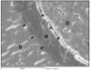
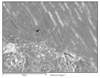
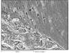
The scanning electron micrographs revealed that removal of the smear layer allowed the adhesive agents and AH26 to enter dentinal tubules. A hybrid-like layer was observed in all the specimens of group 1 and 2 (Fig. 1 and Fig. 2). The dentinal tubules were also open and AH26 could penetrate into the dentinal tubules in Group 3 (Fig. 3). AH26 in group 4 less penetrated into the dentinal tubules than the other groups and no penetration was observed in some area (Fig. 4).
IV. DISCUSSION
Microleakage of oral microorganism through the root filling may be an important cause of failure in root canal therapy10). There are numerous factors that may affect coronal leakage. These include the thickness of the sealer cement, presence of voids within the root canal filling and solubility of the sealer. In addition, the differences in the coefficient of thermal expansion between the obturating materials and dentine must also be considered11). It is important that the sealer cement has low solubility, as dissolution of sealer is one possible cause of leakage subsequent to obturation. In clinical situations, microleakage of bacteria and their by-products is caused by dissolution of root canal sealers and smear layer.
The effect of smear layer on root canal obturation has been evaluated by Kennedy et al.12) who found that its removal improved the sealing of root canals. Epoxy resin-based root canal sealer also enhanced the adaptation of the root canal filling materials to the root dentin especially when smear layer was removed with EDTA3,4). The removal of smear layer improves the adaptation and binding of sealers on root canal walls, especially in the case of AH263). Also in this study, the removal of the smear layer improved the quality of apical sealing. Therefore, removing the smear layer is better for reducing the failure of root canal treatment.
Several studies1,7,8,13) have evaluated the potential of using dentin bonding agents and resins as root canal filling materials. The penetration of the dentin adhesives into the prepared dentin creates a mechanical interlocking and chemical bonds to dentin to create bond strength. Etching of dentin causes demineralization to a depth of several microns and removes the smear layer, thus exposing peri- and intertubular collagen. The primer is hydrophilic in nature facilitating the penetration into the etched dentin, allowing greater surface area coverage and increasing the bond strength14). Assouline et al.15) suggested that dentin adhesives reduced bacterial invasion into dentin after coronal leakage.
The results obtained in this SEM study were consistent with those of Mannocci and Ferrari6). They used two fourth generation dentin bonding agents. They concluded the root canal dentin showed the penetration of resin into the dentinal tubules. But their result of leakage test is different from that of this study. They concluded the apical sealing ability of dentin bonding agents, AH26 and gutta-percha was better when compared with AH26 and gutta-percha in their dye leakage test. But in this study, the root canals filled with gutta-percha and AH26 after the application of dentin adhesive inhibited the passage of F. nucleatum similarly to the canals filled with gutta-percha and AH26 after the removal of smear layer. All-bond 2, fourth generation bonding system and Prime & Bond NT, fifth generation bonding system were used in this study and no significant differences between the two bonding systems were found.
The use of anaerobic bacterial leakage model is more biological and more clinically relevant than dye leakage test, because anaerobic microorganisms are predominant in infections of endodontic origin16). Bae et al.9) found F. nucleatum produced turbidity and/or a color change in this model in 1-2 days and remained viable for 2 weeks and more. Bromcresol purple in bpBHI broth is a chromogenic indicator that changes from purple color at pH 6.8 and above to yellow as the pH decreases to 5.2 in the presence of acidic bacterial by-products9). This indicator was helpful in determining the viability of the test bacteria in the upper chamber as well as aiding in leakage detection. Additional researches for developing dental adhesives in endodontics seem to be necessary to simplify the techniques, to enhance the biocompatibility, and to reduce the microleakage.
V. CONCLUSION
The purpose of this study was to evaluate the sealing ability of epoxy resin-based sealer (AH26) used in conjunction with two different dental adhesives and epoxy resin-based sealer alone with or without smear layer using an anaerobic bacterial leakage model.
The samples of Group 4(AH26 and gutta-percha without treatment of EDTA) leak significantly more than the other three groups (p<0.0005).
There was no statistical significant difference among the other test groups.
Scanning electron microscopic examination of the dentin and the adhesive materials in Group 1 and 2 revealed the presence of characteristic hybrid layer along with microtags of resin penetrating deep into the dentinal tubules.

 XML Download