

 PDF
PDF ePub
ePub Citation
 Print
Print


Abstract
The purpose of this study was to investigate the bonding of resin-based root canal sealer, AH26 when the sealer was applied as a thin layer between dentine and gutta-percha surface.
In this study, forty non-caries extracted human molars and resin-based root canal sealer(AH 26, DeTrey/Dentsply, Germany) were used. Disks of gutta-percha, 6mm in diameter·6mm thick (Diadent/Dentsply, Korea) for thermoplastic obturation were used and dentin surfaces were treated with 2% NaOCl(Group 1) or 2%NaOCl+17% EDTA(Group 3). Disks of gutta-percha, 6mm in diameter·6mm thick (Diadent/Dentsply, Korea) for conventional obturation were used and dentin surface were treated with 2% NaOCl(Group 2) or 2%NaOCl+17% EDTA(Group 4). Enamel was removed by a horizontal section 1mm below the deepest portion of the central occlusal groove by using a water-cooled low speed diamond saw. A second horizontal section was done around cementoenamel junction. Exposed dentin surface was cut to approximately 8×8 mm rectangular shape and was ground against 320, 400, 600 grade silicon carbide abrasive paper serially. After grinding, the dentine surface were soaked in a solution of 2% NaOCl for 30 minutes and twenty of specimens were treated with 17% EDTA solution for 1 minute. The treated specimens were washed and dried. Root canal sealer, AH26 was prepared according to the manufacture's instructions. The Gutta-percha and dentin surface were coated with a thin layer of the freshly mixed sealer. The specimens were left overnight at room temperature. After their initial set, they were transferred to an incubator at 37℃ for 72 h. After 72 hours, resin blocks were made. The resin block was serially sectioned vertically into stick of 1·1mm. Twenty sticks were prepared from each group.
After that, tensile bond strength for each stick was measured with Microtensile Tester. Failure patterns of the specimens at the interface between gutta-percha and dentin were observed under the SEM(×1000) and Stereomicroscope (LEICA M420, Meyer Inst., TX U.S.A) at 1.25 ×25 magnification. The results were statistically analysed by using a One-way ANOVA and Tukey's test.
The results were as follows;
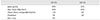
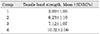
1. Tensile bond strengths(mean±SD) were expressed with ascending order as follows: Group 1, 3.09±1.05MPa ; Group 2, 6.23±1.16MPa ; Group 3, 7.12±1.07MPa ; Group 4, 10.32±2.06MPa.
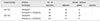
2. Tensile bond strengths of the group 2 and 4 used disks of gutta-percha for conventional obturation were significantly higher than that of the group 1 and 3 used for thermoplastic obturation. (p < 0.05)
3. Tensile bond strengths of the group 3 and 4 treated with 2% NaOCl+17% EDTA were significantly higher than that of the group 1 and 2 treated with 2% NaOCl. (p < 0.05)
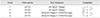
4. In analysis of failure patterns at the interface between sealer and gutta-percha, there were observed 49 (61%)cases of adhesive failure patterns and 31(39%) cases of mixed failures patterns.
Figures and Tables

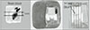
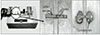
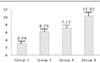
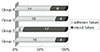
Fig. 6
Failure patterns of the specimens at the interface of the Group1. a,b: Stereomicroscope(1.25×25) c,d:SEM(×1000) S:sealer G:Gutta-percha D:dentin surface.
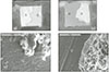
Fig. 7
Failure patterns of the specimens at the interface of the Group2. a,b: Stereomicroscope(1.25×25) c,d:SEM(×1000) S:sealer G:Gutta-percha D:dentin surface.
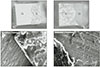
Fig. 8
Filure patterns of the specimens at the interface of the Group3. a,b: Stereomicroscope(1.25×25) c,d:SEM(×1000) S:sealer G:Gutta-percha D:dentin surface.
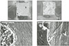
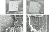
Fig. 9
Filure patterns of the specimens at the interface of the Group4. a,b: Stereomicroscope(1.25×25) c,d:SEM(×1000) S:sealer G:Gutta-percha D:dentin surface.


References
1. Tagger M, Tagger E, Tjan AH, Bakland LK. Measurement of adhesion of endodontic sealers to dentin. J Endod. 2002. 28:351–354.

2. Wennberg A, Orstavik D. Adhesion of root canal sealers to bovine dentine and gutta-percha. Int Endod J. 1990. 23:13–19.
3. Lee KW, Williams MC, Camps JJ, Pashley DH. Adhesion of endodontic sealers to dentin and gutta-percha. J Endod. 2002. 28:684–688.
4. Gettleman BH, Messer HH. Adhesion of sealer cements to dentin with and without the smear layer. J Endod. 1991. 17:15–20.
5. Kataoka H, Yoshilka T, Suda H. Dentin bonding and sealing ability of a new root canal resin sealer. J Endod. 2000. 26:230–235.
6. Calt S, Serper A. Time-dependent effects of EDTA on dentin structures. J Endod. 2002. 28:17–19.
7. Timpawat S, Harnirattisai C. Adhesion of a glass-ionomer root canal sealer to the root canal wall. J Endod. 2001. 27:168–171.
8. Pashley DH, Carvalho RM, Sano H, Nakajima M. The microtensile bond test : a review. J Adhes Dent. 1999. 1:299–309.
9. Armstrong SR, Boyer DB. Microtensile bond strength testing and failure analysis of two dentin adhesives. Dent Mater. 1998. 14:44–50.
10. Grossman LI, Oliet S, Del Rio CE. Endodontic Practice. 11th edn. Philadelphia: Lea & Febiger;255.
11. Goldman M, Goldman LB, Cavaleri R. The efficacy of several endodontic irrigating solutions : a scanning electron microscopic study. part. 2. J Endod. 1982. 8:487–492.
12. Wiener BH, Schilder H. A comparative study of important physical properties of various root canal sealersII Evaluation of dimensional changes. Oral Surg Oral Med Oral Pathol. 1971. 32:928–937.
13. De Gee AJ, Wu MK, Wesselink PR. Sealing properties of Ketac Endo glass ionomer cement and AH26 root canal sealers. Int Endod J. 1994. 27:239–244.
14. Jeffrey IW, Saunders WP. An investigation into the bond strength between a root canal sealer and root-filling points. Int Endod J. 1987. 20:217–222.
15. Orstavik D, Eriksen HM, Beyer-Olsen EM. Adhesive properties and leakage of root canal sealers in vitro. Int Endod J. 1983. 16:59–63.
16. Pashley DH, Sano H, Ciucchi B, Yoshiyama M. Adhesion tesing of dentin bonding agents : A review. Dent Mater. 1995. 11:117–125.
17. Zmener O, Spielberg C, Lamberghini F, Rucci M. Sealing properties of a new epoxy resin-based root-canal sealer. Int Endod J. 1997. 30:332–334.
18. Marciano J, Michailesco PM. Dental gutta-percha : Chemical composition ,X-ray identification, enthalpic studies, and clinical implications. J Endod. 1989. 15:149–153.
19. Branstetter J, von Fraunhofer JA. The physical properties and sealing action of endodontic sealer cements : a review. J Endod. 1982. 8:312–316.
20. Schafer E, Olthoff G. Effect of three different sealers on the sealing ability of both thermafil obturators and cold laterally compacted gutta-perch. J Endod. 2002. 28:638–642.
21. Sano H, Shono T, Sonoda H, Takatsu T, Ciucchi B, Carvalho R, Pachley DH. Relationship between surface area for adhesion and tensile bond strength-Evaluation of a microtensile bone test. Dent Mater. 1994. 10:236–240.
22. De Gee AJ, Wu M-K, Wesselink PR. Sealing properties of Ketac-Endo glass ionomer cement and AH26 root canal sealers. Int Endod J. 1994. 27:239–244.
23. Sousa-Neto MD, Marchesan MA, et al. Effect of Er:YAG laser on adhesion of root canal sealers. J Endod. 2002. 28:185–187.
24. Baumgartner JC, Mader CL. A scanning electron microscopic evaluation of four root canal irrigation regimens. J Endod. 1987. 13:147–157.
25. Azar NG, Heidari M, et al. In vitro cytotoxicity of a new epoxy resin root canla sealer. J Endod. 2000. 26:462–465.
26. Lalh MS, Titley K, Torneck CD, Friedman S. The shear bond strength of glass ionomer cement sealers to bovine dentin conditioned with common endodontic irrigants. Int Endod J. 1999. 32:430–435.
27. Meryon SD, Tobias RS, Jakeman KJ. Smear removal agents : a quantitative study in vivo and in vitro. J Prosthet Dent. 1987. 57:174–179.
28. Cohen Stephen, Burns Richard C. Pathways of the pulp. seventh edition. 508–509.
29. Jang SW, Hong CE. Chi-yuleul wihan geungwanchilyo Atlas.
 XML Download