

 PDF
PDF ePub
ePub Citation
 Print
Print


Abstract
Object
The purpose of this study were to evaluate the microtensile bond strength of resin fiber reinforced post to radicular dentin using resin cement according to various dentin surface treatment and to observe the interface between post and root dentin under SEM.
Material and Method
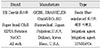
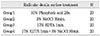
A total 16 extracted human single rooted teeth were used.
A lingual access was made using a #245 carbide bur in a high-speed handpiece with copious air water spray. The post space was mechanically enlarged using H-file(up to #60) and Gates Glidden bures(#3). This was followed by refining of the canal space using the calbrating drill set provided in ER Dentinpost(GEBR, BRASSELER GmbH&Co. KG).
The 16 teeth were randomly distributed into 4 group of 4 teeth.
Group 1 teeth had their post space prepared using 10% phosphoric acid as root canal surface treatment agent during 20s. The canal was then rinsed with saline and dried with paper point.
Group 2 teeth had their post space prepared using 3% NaOCl as root canal surface treatment agent during 30min. The canal was then rinsed with saline and dried with paper point.
Group 3 teeth had their post space prepared using 17% EDTA as root canal surface treatment agent during 1min. The canal was then rinsed with saline and dried with paper point.
Group 4 teeth had their post space prepared using 17% EDTA as root canal surface treatment agent during 1min. After rinsing with saline, the canal was rinced 10ml of 3% NaOCl for 30min.
After drying with paper point, the post(ER Dentinpost, GEBR, BRASSELER GmbH&Co. KG) was placed in the treated canals using resin cement. Once the canal was filled with resin cement(Super bond C&B sunmedical co. Ltd.), a lentulo was inserted to the depth of the canal to ensure proper coating of the root canal wall.
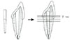
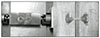
After 24 hours, acrylic resin blocks(10·10·50mm) were made. The resin block was serially sectioned vertically into stick of 1·1mm. Twenty sticks were prepared from each group. After that, tensile bond strengths for each stick was measured with Microtensile Tester.
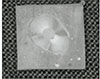
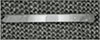
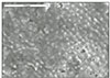
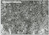
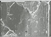
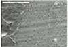
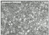
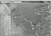
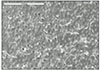
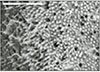
Failure pattern of the specimen at the interface between post and dentin were observed under SEM.
Results
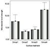
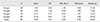
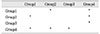
1. Tensile bond strengths(meen±SD) were expressed with ascending order as follows ; group 4, 12.52±6.60 ; group 1, 7.63±5.83 ; group 2, 4.13±2.31 ; group 3, 3.31±1.44.
2. Tensile bond strengths of Group 4 treated with 17% EDTA +3%NaOCl were significant higher than those of group 1, 2 and 3 (p<0.05).
3. Tensile bond strengths of Group 1 treated with 10% phosphoric acid were significant higher than those of group 2 (p<0.05). Tensile bond strengths of Group 4 treated with 17% EDTA +3% NaOCl was significant higher than those of other groups.
Figures and Tables


References
1. Martinez-Insua A, da Silva L, Rilo B, Santana U. Comparision of the fracture resistance of pulpless teeth restored with a cast post and core or carbon-fiber post with a composite core. J Prosthet Dent. 1998. 80:527–532.

2. Asmussen E, Peutzfeldt A, Heitmann T. Stiffness, elastic limit, and strength of newer types of endodontic posts. J Dent. 1999. 27:275–278.
3. Platt JA. Resin-based luting cements. Compend Contin Educ Dent. 2000. 21:740–742.
4. Calt S, Serper A. Time-Dependent effects of EDTA on dentin structures. J Endod. 2002. 28:17–19.
5. Harrison JW, Hand RE. The effect of dulution and orgnic matter on the antibacterial of 5.25% sodium hypochlorite. J Endod. 1981. 7:128–132.
6. Morris MD, Lee KW, Agee KA, Bouillaguet S, Pashley DH. Effects of sodium hypochlorite and RC-prep on bond strengths of resin cement to endodontic surfaces. J Endod. 2001. 27:753–757.
7. Gaston BA, West LA, Liewehr FR, Fernandes C, Pashley DH. Evaluation of regional bond strength of resin cement to endodontic surfaces. J Endod. 2001. 27:321–324.
8. Pashley DH, Carvalho RM, Sano H, Nakajima M, Yoshiyama M, Shono Y, Fernandes CA, Tay F. The microtensilebondtest :a review. J Adhes Dent. 1999. 1:299–309.
9. Sano H, Shono T, Sonoda H, Takatsu T, Ciucchi B, Carvalho R, Pashley DH. Relation between surface area for adhesion and tensile bond strength -Evaluation of a microtensile bond test. Dent Mater. 1994. 10:236–240.
10. Pashley DH, Samo H, Ciucchi B, Yoshiyama M, Carvalho RM. Adhesion testing of dentin bonding agents : A Review. Dent Mater. 1995. 11:117–125.
11. Chappell R, Schreiner R, Glaros A, Eixk J. Pilot study to determine sample size for micro-tensile testing. J Dent Res. 1997. 76:38.
12. Zheng L, Pereira PN, Nakajima M, Sano H, Tagami J. Relationship between adhesive thickness and microtensile bond strength. Oper Dent. 2001. 26:97–104.
13. Sudsangiam S, Van Noort R. Do dentin bond strength tests serve a useful purpose? J Adhes Dent. 1999. 1:57–67.
14. Van Noort R, Noroozi S, Howard IC, Cardew G. A critique of bond strength measurements. J Dent. 1989. 17:61–67.
15. Ferrari M, Vichi A, Grandini S. Efficacy of different adhesive techniques on onding to root canal walls: an SEM investigation. Dent Mater. 2001. 17:422–429.
16. Chow TW. Mechanical effectivenss of root canal irrigation. J Endod. 1983. 9:475–479.
17. Scelza MF, Antoniazzi JH, Scelza P. Efficacy of final irrigation-a scanning electron microscopic evaluationcccc. J Endod. 2000. 26:355–358.
18. Cunningham WT, Balekjian AY. Effect of temperature on collagen-dissolving ability of sodium hypochlorite endodontic irrigant. Oral Surg Oral Med Oral Pathol. 1980. 49:175–177.
19. Niu W, Yoshioka T, Kobayashi C, Suda H. A scanning electron microscopic study of dentinal erosion by final irrigation with EDTA and NaOCl solutions. Int Endod J. 2002. 35:934–939.
20. Villegas JC, Yoshioka T, Kobayashi C, Suda H. Obturation of accessory canals after four different final irrigation regimes. J Endod. 2002. 28:534–536.
21. Serper A, Calt S. The demineralizing effects of EDTA at different concentrations and pH. J Endod. 2002. 28:501–502.
22. Kitasako Y, Burrow MF, Tnikaido T, Tagami J. Long-term tensile bond durability of two different 4-MATA containing resin cement to dentin. Dent Mater. 2002. 18:276–280.
23. Goldman M, Devitre R, white R, Nathanson D. An SEM study of post cemented with an unfilled resin. J Dent Res. 1984. 63:1003–1005.
24. Chang JC, Hurst TL, Hart DA, Estey AW. 4-META use in dentistry: a literature review. J Prosthet Dent. 2002. 87:216–224.
25. Calt S, Serper A. Smear layer removal by EGTA. J Endod. 2000. 26:459–461.
26. Mak YF, Lai SC, Cheung GS, Chan AW, Tay FR, Pashley DH. Micro-tensile bond testing of resin cements to dentin and an indirect resin composite. Dent Mater. 2002. 18:609–621.
27. Vichi A, Grandini S, Davidson CL, Ferrari M. An SEM evaluation of several adhesive systems used for bonding fiber posts under clinical conditions. Dent Mater. 2002. 18:495–502.
 XML Download