

 PDF
PDF ePub
ePub Citation
 Print
Print


Abstract
This study was done to present a criterion in selection of the most proper light sources and materials by measuring metamerism index(MI) of the light curing composite resins with spectrocolorimeter. Metamerism is defined when two objects appear to be the same color in one illuminant but different in another. This is due to the fact that they have different spectral curves that fail to match under the second illuminant.
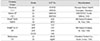
In this study, A1 & A3 shade of five light curing composite resins (Esthet-X, Filteck Z250, Filteck A110, Charisma, Vitalescence) were chosen based on Vita shade. Five samples were made for shade of each product with Teflon mold (diameter: 15mm, thickness: 2mm).
Metamerism index of each samples on a Barium sulfate plate (L*=96.54, a*=0.19, b*=0.01) prepared for sample fixation were measured with spectrocolorimeter(Miniscan XE plus, Model 4000s, Hunter Lab, USA) by applying standard light source D65, C, Fcw, TL84 and A. Standardization was done with reference standard (X=80.8, Y=85.7, Z=90.8) and light trap. The results were as follows.
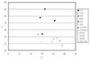
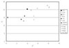
Different resins with same Vita shade showed recognizable color difference(ΔE*>2).
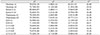
All composites had MI below accepted value 0.5 between standard illuminant(D65, C, & A) and below 1.5 under fluorescent condition (Fcw & TL84).
MI value between D65 and A showed higher value than MI value between other source of light(p<0.01).
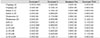
All resins except Z250 showed MI value that A3 is higher than A1 between D65 and A(p<0.05).
Figures and Tables
References
1. Clark EB. An analysis of tooth color. J Am Dent Assoc. 1931. 18:2093–2103.

2. Clark EB. Tooth color selection. J Am Dent Assoc. 1933. 20:1065–1073.
3. Hayashi T. Medical color standard. V. Tooth crown. 1967. Tokyo: Japan Color Research Institute.
4. Miller LL. Organizing color in dentistry. J Am Dent Assoc. 1987. 12. Spec No:26E–40E.
5. Johnston WM, Kao EC. Assessment of appearance match by visual observation and clinical colorimeter. J Dent Res. 1989. 68(5):819–822.
6. Hwang IN, Oh WM. Colorimetric Analysis of Extracted Human Teeth and Five Shade Guides. J Korean Acad Conserv Dent. 1997. 22(2):769–781.
7. Sproull RC. Color matching in dentistry. Part II: Practical applications of the organization of color. J Prosthet Dent. 1973. 29:556–566.
8. Goodkind RJ, Keenan KM, Schwabacher WB. A comparison of Chromascan and spectrophotometric color measurement of 100 natural teeth. J Prosthet Dent. 1985. 53:105–109.
9. Schwabacher WB, Goodkind RJ. Three-dimensional color coordinates of natural teeth compared with three shade guide. J Prosthet Dent. 1990. 64(4):425–431.
10. Yeh CL, Powers JM, Miyagawa Y. Color of selected shades of composites by reflection spectrophotometry. J Dent Res. 1982. 61(10):1176–1179.
11. Cho KM, Shin DH. Color Analysis of the Natural Teeth with a Modified Intraoral Spectrophotometer. J Korean Acad Conserv Dent. 1998. 23(1):223–235.
12. Lee MY, Shin DH. New Evaluation Technique in Teeth Color Using Digital Camera. J Korean Acad Conserv Dent. 1997. 23(1):325–333.
13. Kim HS, Um JM. A study on color differences between composite resins and shade guides. J Korean Acad Conserv Dent. 1996. 21(1):107–120.
14. Cho KY, Hwang IN, Choi HR, Oh WM. Comparative Evaluation of Light-cured Composite Resins Based on Vita Shade by Spectrocolorimeter. J Korean Acad Conserv Dent. 1998. 23(1):424–432.
15. Hwang IN, Lee KW. Translucency of Light Cured Composite Resins Depends on Thickness & Its Influence on Color of Restorations. J Korean Acad Conserv Dent. 1999. 24(4):604–613.
16. Swepston JH, Miller LL. Esthetic matching. J Prosthet Dent. 1985. 54:623–625.
17. Robertson AR. Colorimetry. Rep Prog Phys. 1978. 41:471–510.
18. Thornton WA. How strong metamerism disturbs color spaces. Color Res Appl. 1998. 23(6):402–407.
19. MiniScan XE plus users guide. HunterLab.
20. Park EJ. The Basic of Color Modeling. 1996. 2nd ed. Seoul: Mijinsa;56–194.
21. Park DY. Practical Chromatology. 1992. enlarged ed. Seoul: Bando Publisher;99–120.
22. The Korean Industrial Standard, KS A 0065.
23. Grajower R, Revah A, Sorin S. Reflectance spectra of natural and acrylic resin teeth. J Prosthet Dent. 1976. 36(5):570–579.
24. Seghi RR, Hewlett ER, Kim J. Visual and instrumental colorimetric assessments of small color differences on translucent dental porcelain. J Dent Res. 1989. 68:1760–1764.
 XML Download