

 PDF
PDF ePub
ePub Citation
 Print
Print


I. Introduction
Complete obturation of the root canal with an inert filling material and creation of a hermetic apical seal have been proposed as goals for successful endodontic treatment. Currently, the methods most frequently used in filling root canals involve the use of core points, such as gutta-percha, inserted in conjunction with sealers or pastes. Many types of root canal sealers using zinc oxide-eugenol, resins, calcium hydroxide, glass ionomer, calcium phosphate and so on have been commercially available and clinically used. Since these materials will be in direct contact with periapical tissues for a long period of time, their biocompatibility is important.
It is well known that calcium phosphate biomaterials show high stability and low irritation to tissues, but it does not set itself by any chemical reaction. Thus, it might be unsuitable for use as a major component of a root canal sealer. On the other hand, according to Brown and Chow1), a certain combinations of finely powdered calcium phosphates, when mixed with water, harden into cements which have similar chemical composition and crystal structures to those of tooth and bone mineral. That is a self-setting calcium phosphate cement (CPC). For example, the combination of tetracalcium phosphate (TTCP) and dicalcium phosphate anhydrous (DCPA) react to form hydroxyapatite (HA) which is biocompatible and osteoconductive. Both the setting ability and biocompatibility made CPC as a potentially useful material in a variety of dental and medical application. In vitro studies2-4) and an animal model5) have indicated that it was useful in endodontics as a sealer in root canal treatment. In addition to being used as a sealer, some studies have shown that CPC could also seal a furcation perforation6) and could used as an apical barrier for apexification7) and direct pulp-capping agent8). These results suggest that CPC has potential to promote the healing of bone in endodontic treatment.
Apatite Root Sealers (ARS) are α-tricalcium phosphate-based root canal sealers. Biocompatibility studies on ARS have been conducted by several researchers9-11). Efforts for histopathological evaluation on these materials seem to contradict each other9,10). Bilginer et al.9) reported a mild to moderate inflammatory reaction to ARS, whereas Yoshikawa et al.10) reported a severe inflammatory reaction. Telli et al.11) evaluated the cytotoxicity of ARS with MTT assay and reported ARS did not exert any cytotoxic effects. But cytotoxicity change of ARS by setting reaction was not yet reported. The main objective of this study was to evaluate time-dependent changes in cytotoxicity of ARS Type I, II and III in comparison with those of four other commonly used endodontic sealers (Pulp Canal Sealer EWT, AH Plus, Sealapex and Ketac Endo).
II. Materials and methods
Test materials and sample preparation
The test materials used were Apatite Root Sealer (ARS) Type I, II and III (Sankin kogyo, Tokyo, Japan), Pulp Canal Sealer EWT (Kerr, Detroit, MI, USA), AH Plus (DeTrey Dentsply, Zurich, Switzerland), Sealapex (Kerr, Detroit, MI, USA) and Ketac Endo (Espe, Seefeld, Germany). The compositions of ARS Type I, II and III were listed in Table 1. The root canal sealers were prepared under aseptic conditions according to the manufacturer's directions and filled into sterile glass rings (5mm inner diameter and 4mm high). The filled glass ring was placed in a well of 24-well plates (NUNC, Denmark) containing 2ml of complete culture medium. Plates were then kept at 37℃ in a humidified 5% CO2-containing incubator. Extractions were obtained after mixing of sealers during the following 7 observation periods of time :
At the end of each observation period, the glass rings were removed and washed with a fresh culture medium and transferred to a next new wells containing 2ml of complete culture medium and incubated for the next time interval. The wells containing glass rings with no sealer were served as negative controls. The sealer extracts were collected into syringes and filtered with a 0.2µm sterile syringe filter (Corning, Germany) to ensure sterile conditions.
Cell culture
L929 mouse fibroblasts were cultured in Minimum Essential Medium (MEM; Gibco-BRL, USA) supplemented with 100U/ml of penicillin, 50µg/ml of streptomycin (Gibco-BRL, USA), and 10% fetal bovine serum (FBS; Gibco-BRL, USA) at 37℃ in a humidified atmosphere of 5% CO2. The cells were plated into 96-well flat bottom plates (Costar, USA) at a density of 15,000 cells/100µl in each well and incubated for 24h to allow attachment. The complete culture medium was replaced with 100µl of sealer extract and control. Eight wells were used for single sealer extract from each time interval and control. After 24h incubation, the cytotoxicity was accessed.
MTT assay
The MTT assay was carried out according to Mosmann12). This assay focused on the capacity of mitochondrial dehydrogenase enzymes in living cells to convert the yellow water-soluble tetrazolium salt (3-[4,5-dimethyl thiazol-2-yl]-2,5-diphenyl tetrazolium bromide) (MTT) into dark-blue insoluble formazan crystals. The amount of formazan generated was directly proportional to the cell number. 20µl of a solution of 5mg/ml MTT (Sigma Chemical Co., USA) in phosphate buffered saline were added to each well. The cells were incubated for 3h at 37℃ and the plates were inverted and blotted on filter paper to remove excess MTT medium. 100µl dimethyl sulfoxide (DMSO; Sigma Chemical Co., USA) were added to solubilize the formazan product and the plates were shaken. Optical densities (OD's) were measured at 570nm using ELISA READER (Thermomax, Molecular device, USA).
Neutral red(NR) assay
The neutral red (NR) assay was performed basically as the method reported by Borenfreund and Puerner13). In this assay, cell viability is indicated by the uptake of the vital dye, neutral red, into lysosomes of viable cells. A 0.33% neutral red (Sigma Chemical Co., USA) was diluted to 50µg/ml in complete culture medium. 200µl of the staining solution were added to each well after removal of the sealer extract. The cells were incubated for 3h at 37℃. All wells were aspirated and the cells fixed with 100µl 1% calcium chloride w/v in 0.5% formaldehyde. All wells were aspirated again and destained with 100µl 1% acetic acid in 50% ethanol. The plates were shaken and optical densities (OD's) were measured at 540nm using ELISA READER (Thermomax, Molecular device, USA).
Data processing and statistical analysis
The percentage of cytotoxicity of each experimental group was calculated using the following formula:
% cytotoxicity = (1- mean OD of experimental group/mean OD of control group)×100
The whole procedure was repeated five times for each extract, using eight replicates of each extract per experiment. The statistical analysis was obtained by the Kruskal-Wallis test and the Friedman test. Differences were considered to be significant at a p-value of less than 0.05.
III. Results
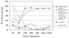
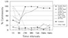
The results of the MTT assay are demonstrated in Table 2 and Figure 1 and results of the NR assay are demonstrated in Table 3 and Figure 2. Apatite Root Sealer (ARS) Type I, II and III represented slight cytotoxicity (0.55 - 23.65%) at all observation periods in both assays. There was no significant difference in the cytotoxic effects among ARS Type I, II and III at each time interval (p>0.05). AH Plus and Ketac Endo induced significant cytotoxic effects at early stage (p<0.05) but slight cytotoxicity after 8h (AH Plus) and 24h (Ketac Endo). Pulp Canal Sealer EWT and Sealapex showed moderate cytotoxicity at early stage (1h, 8h) but sever, long-lasting cytotoxicity until 4wks (p<0.05). In most cases, cytotoxic effects of each sealer extract showed similar patterns between the MTT assay and the NR assay.
IV. Discussion
Biocompatibility of dental materials can be evaluated either in vivo or in vitro. The in vitro experimentation is simple, reproducible, inexpensive, relevant, and suitable for initial screening of the material's cytotoxicity, and it also precludes the use of experimental animals14). Many methods have been proposed to assess in vitro the cytotoxicity of dental materials. In this study, the reduction of MTT by active cells and the uptake of neutral red into lysosomes were measured using two viability assays. These assays can be performed very effectively; only few cells are needed for rapid, reliable and inexpensive screening purposes of a large number of samples in a short time. Automated processing with a microplate reader and subsequent automated analyses of original data are additional advantages of the systems15). The MTT assay and the NR assay exhibited similar patterns for most test materials in the present study.
The actual availability of potentially toxic substances from sealers differs greatly, depending on their setting reactions and at which stage they are tested14). In the present study, cytotoxicity was evaluated for each sealer extract obtained from various time intervals (immediately after mixing, during setting and after setting of sealers), enabling assessment of early and late cytotoxic effects of the sealers.
Pulp Canal Sealer EWT, a zinc oxide eugenol-based sealer, showed moderate cytotoxicity immediately after mixing, but thereafter its cytotoxicity began to increase and remained at a high level until 4wks. The cause of this long-lasting cytotoxicity may be related to unreactive eugenol or zinc ion remaining in the zinc oxide and eugenol mixture16-18). Similar results were obtained in the studies of ZOE by Others17,18).
Sealapex also showed severe long-lasting cytotoxicity until the end of the observation periods after it initially showed moderate cytotoxicity. This toxicity is related to strong alkalinity of the calcium hydroxide material that changes the medium color19-21). Sealapex was unstable in an aqueous environment, so the hardened sealer was not completely hardened and collapsed in the medium. This might be attributed to the capability of calcium hydroxyl ions to diffuse and precipitate from the set sample in the medium. Previous experiment done by Duarte et al.22) reported about the ability of calcium and hydroxyl ion release.
AH Plus, a new formulation of the epoxy resin-based sealer AH26, exhibited severe cytotoxicity immediately after mixing, but its cytotoxicity sharply decreased after 8h. These results were in accordance with the study by Azar et al.17). Leyhausen et al.23) reported that AH Plus caused minor or no cytotoxic effect and no genotoxic effect with 24h set sealer. Short time cytotoxicity of AH Plus could probably be due to its low contents of water-soluble toxic substances (presumably formaldehyde and epoxy derivatives of bisphenol-A-diglycidylether) and short setting time (8h)17).
Ketac Endo, glass ionomer based-sealer, induced high cytotoxic effect at the early stage like the AH Plus, but it showed slight cytotoxicity after 24h. Its early cytotoxicity may be due to high acidity at the freshly mixed stage. According to Ersev et al.24), Ketac-Endo had little cytotoxic effect on L929 cells after a setting time of 24h and 1wk. Beltes et al.25) also reported that very low cytotoxicity was observed after setting of sealers
In this experiment, ARS Type I, II and III, calcium phosphate based-sealers, showed very low cytotoxicity from the early stage to the late stage in both assays. Although ARS Type II and Type III contain iodoform, the difference among the cytotoxicity of ARS Type I, II, and III was not statistically significant (p>0.05). This results appear to be in agreement with a previous study by Telli et al.11). In that study, ARS Type I, II and III did not exert any cytotoxic effects with sealer extracts obtained during 1-2h after mixing using the MTT assay.
Although in vitro assays may not correlate highly with in vivo data, there were two histopathological studies on these materials that seem to contradict each other9,10). Bilginer et al.9) reported a mild to moderate inflammatory reaction to ARS, and suggested that ARS Type II and III were found more biocompatible than ARS Type I. Whereas Yoshikawa et al.10) reported a severe inflammatory reaction to ARS Type I. According to Mjor et al.26), the surface properties and geometry of the material in contact with the tissue may be of importance, and bacterial contamination of the wound area may give rise to an inflammatory process of greater magnitude than any material reaction. This criteria must always be taken into consideration when evaluating histopathological studies.
A great many researchers investigated calcium phosphate materials as a root canal sealers2-5,27). Calcium phosphate cement(CPC) has the following advantages over the conventionally used sealers: (a) Its high biocompatibility suggests that inadvertent extrusion beyond the apical foramen should be well tolerated by the periapical tissues. (b) CPC would be tolerated by patients who might be allergic to eugenol. (c) It appears to be devoid of dimensional changes during setting and provides superior adaptation to the canal surface, thus allowing a better hermetic seal of the apical foramen and accessory canals located in the apical third of the root. (d) Moisture in the canal will not decrease the strength of CPC; the mechanical strength of CPC actually increased when placed in saliva or plasma-like fluids. (e) The setting time of CPC can be controlled easily to provide sufficient working time.
By the way, CPC does not compare favorably with currently available dental cements in terms of setting time and strength. To overcome the shortcomings of CPC, it must be modified in its composition and should be admixed with various addictives. But this alteration can have an influence on other properties, although some properties are improved, therefore to suit clinical need as an endodontic materials, it is necessary to be further studied.
V. Conclusions
This study evaluated the cytotoxicity of calcium phosphate root canal sealers using the MTT assay and the NR assay.
ARS Type I, II and III showed low cytotoxicity at all observation periods, and proved to be biocompatible materials.
In most cases, cytotoxic effects of each sealer extract showed similar patterns between the MTT assay and the NR assay, so both assays were useful cytotoxicity tests.
Calcium phosphate root canal sealers need further evaluation and development.


 XML Download