

 PDF
PDF ePub
ePub Citation
 Print
Print


I. INTRODUCTION
One of the goals in root canal instrumentation is to clean and shape the root canal system to form a continuously tapered preparation from the coronal access to the apical foramen while maintaining the original shape and position of the apical foramen1). Cleaning and shaping of root canal is the most important step in endodontic treatment. However, this is difficult to achieve with conventional stainless-steel instruments in narrow and curved root canals2).
Root canal anatomy has great varieties3) and has been reported to have curves beginning at almost any level. Even canals that are apparently straight may have curvatures and irregularities in the apical one-third4). In curved canals, there is a tendency for all preparation techniques to transport the prepared canal away from the original axis and every file has a tendency to straighten the curved canal, resulting in excessive removal of the dentin of the outer wall of the apical curvature and the formation of teardrop-shaped foramen2). Roane5) referred this inherent characteristic for endodontic files as the "restoring force" of the instrument.
To overcome the limitations of stainless-steel instruments, endodontic instruments made of the super-elastic alloy nickel-titanium (Ni-Ti) have been introduced. Walia et al.6) found that Nitinol files had two to three times more elastic flexibility in bending on torsion, as well as superior resistance to torsional fracture, compared with similar sized stainless-steel files. Favorable effects on canal shape have been noted with the use of Ni-Ti instruments for canal preparation. Root canal prepared with Ni-Ti instruments stayed more centered in the canal, produced rounder preparations, and reduced transportation and ledging7). Serene et al.8) found that preparations using Ni-Ti instruments require less force and at least 20% less preparation time.
Ni-Ti rotary endodontic instruments place new demands on instruments used to clean and shape root canals. Recently, Ni-Ti rotary instruments have become a mainstay for not only endodontists but also general practitioners. And various hybrid techniques are introduced for faster and more effective cleaning and shaping of root canal. By incorporating a hybrid technique that combines variable tapers, different systems and modification of individual techniques, many of the endodontics problems that had arisen during the early years of rotary instrumentation have become noticeably reduced8).
Buchanan9) found that Ni-Ti rotary files were safe, fast, simple to master, and offered predictable cleaning and obturation outcomes, even in novice operators. Manufacturers said that Ni-Ti file systems were easy and fast methods for root canal shaping to all operators. However, Mandel et al.10) reported that novice operators need a learning period when using the Ni-Ti file system, and there were few studies for efficiency of Ni-Ti rotary files in novice operators.
The goal of this study was to evaluate the efficiency of the Ni-Ti rotary files in curved canals shaped by novice operators and to know which technique is suitable for novice operators.
II. Materials and Methods
Simulated canals
Seventy two transparent resin blocks with simulated root canal (Dentsply Maillefer, Ballaigues, Switzerland) were used for this study. Schneider's method was used to determine the curvature of canals and average curvature was 41.4 degree. All canals were 19.5 mm, consisting of 12 mm straight coronal region, and a 7.5 mm curved apical region. The working length was established with an ISO size 10 instruments by endodontists.
Instrumentation
The seventy two resin blocks were divided equally into three groups: Group 1 canals were instrumented using the ProFile system (Dentsply Maillefer, Ballaigues, Switzerland), Group 2 canals were instrumented using the ProTaper system (Dentsply Maillefer, Ballaigues, Switzerland), and Group 3 canals were instrumented using a Hybrid instrumentation procedure employing both the ProFile and ProTaper systems. Each group of 24 resin blocks were further evenly subdivided into two subgroups; Subgroup A canals were instrumented by four endodontists who had extensive experience and practice with both of the Ni-Ti file systems; Subgroup B canals were instrumented by 4 undergraduate dental school students who had no prior experience with either of the Ni-Ti file systems, each of these undergraduate students was only given a thirty-minute tutorial and demonstration on the specific rotary Ni-Ti file system that they would be using prior to their own experimental instrumentation. Subgroup A root canals was instrumented by the same four endodontists for all three groups (Group 1, Group 2 and Group 3), while Subgroup B root canals were instrumented by 12 undergraduate students from the College of Dentistry at Kangnung National University (Kangnung, Korea)(Table 1).
Files were placed in an electric motor (Aseptico model AEU-20 ITR, Aseptico inc., Woodinville, WA, USA) with a 1:8 reduction contra-angle handpiece (Anthogyr, Sallanches, France). The speed of the file was set at 300 rpm and the torque was set at level 2.
Group 1
Group 1 was instrumented with ProFiles using the crown-down technique recommended by the manufacturer. The ProFiles were used in the following sequence. For coronal shaping, serial use of Orifice shaper #3, #2 to the coronal one-half of the canal, and ProFiles .06/#25, .06/#20, .04/#25 to the coronal two-thirds of the canal were performed in crown-down technique. For apical shaping, ProFile .04/#20 and .04/#25 were sequentially used to the working length(Table 2).
Group 2
Group 2 was instrumented with ProTaper files using the crown-down technique recommended by the manufacturer. The ProTaper files were used in the following sequence. Serial uses of ProTaper S1, Sx to the coronal two-thirds of the canal, and ProTaper S1, S2 to the working length were performed in crown-down technique. For apical shaping, ProTaper F1, F2 were sequentially used to the working length(Table 2).
Group 3
Group 3 was instrumented by Hybrid method of ProTaper files in combination with ProFiles. Serial uses of ProTaper S1, Sx to the coronal two-thirds of the canal, and ProTaper S1, S2 to the working length were performed in crown-down technique. For apical shaping, ProFile .04/#20 and .04/#25 were sequentially used to the working length(Table 2).
All canals were copiously irrigated with saline before preparation and after use of each instrument using a disposable syringe with an endodontic irrigation needle (Max-i-Probe, Dentsply, Franklin Park, IL, USA). Prior to use, each canal was soaked in RC-Prep (Premier Dental Products Co., Philadelphia, PA, USA) as a lubricant. Each file was used in one canal only and discarded after use.
Imaging system
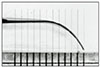
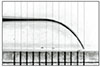
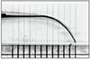
All the canals were stained; methylene blue dye was introduced into the canal to obtain a clear pre-instrumentation image and, all the canals were filled with Vitapex™(Neo Dental Chemical Products Co., Ltd., Tokyo, Japan) for the post-instrumentation image.
Images of all the simulated canals were obtained before and after instrumentation, using digital photography (FinePix S1-Pro, 340 Mega pixel, Fuji Photo Film Co., Ltd., Tokyo, Japan) with 120 mm Medical Nikkor (Nikon Corporation, Tokyo, Japan) macro lens. A mounting device was developed and used to locate the camera and the resin blocks at the same position accurately.
Assessment of canal preparation
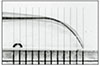
Measurements were made at 7 different levels: 1, 2, 3, 4, 5, 6, and 7 mm from the apical foramen. At each level, 3 measurements were made: post-instrumentation canal diameter, change in outer canal width, and change in inner canal width. Apical transportation and the time taken for each technique, including time for instrument changes and irrigation, were also noted.
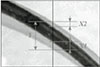
The amount of canal center movement and mean centering ratio were measured by using the greatest distance between the edge of each post-instrumented canal and the corresponding edge of the pre-instrumented canal. The mean centering ratio (CR) was a measure of the ability of the instrument to stay centered in the canal; the smaller the ratio, the better the instrument remained centered in the canal. This ratio was calculated for each instrumented canal using the following formula11): CR = (X1-X2)/Y, as reported by Calhoun and Montgomery12), where X1 is the maximum extent of canal movement, X2 is the movement in the opposite direction, and Y is the diameter of the post-instrumented canal (Fig. 7). The direction of maximum canal transportation, as determined by the direction in which X1, X2, and Y were measured was recorded13).
For statistical analysis, Mann-Whitney U-test was performed to compare the post-instrumentation canal diameters, movements of canal center, the centering ratios and the time taken between operators for each groups by SPSS 10.0 for Windows (SPSS Inc., Chicago, IL, USA).
III. RESULTS
Changes in Post-Instrumentation Canal Diameter
The mean value of post-instrumentation canal diameters and statistical analysis with Mann-Whitney U-test are shown in Table 3 and Table 4. In all Groups, post-instrumentation canal diameters in novice operators were greater than those in endodontists.
Post-instrumentation canal diameters at 1, 2, 5, 6, 7 mm level in ProFile, 6 mm level in ProTaper, and 3 mm level in Hybrid showed a significant difference between operators (P<0.05).
Movements of Canal Center
The mean value of movements of canal center and statistical analysis with Mann-Whitney U-test are shown in Table 5 and Table 6. There were movements of canal center in all groups. Movements of canal center in novice operators were greater than those in endodontists at 1, 2, 4, 5 mm level in ProFile, at 1, 6 mm level of ProTaper and at 1, 2, 3, 5, 6, 7 mm level in Hybrid. Movements of canal center at 5 mm level in ProFile and at 3 mm level in Hybrid showed a significant difference between operators (P<0.05). In all groups, canal center moved towards the inner portion of curvature at 4, 5, 6, 7 mm level from apex and moved towards the outer portion of curvature at 1, 2, 3 mm level. However, at 3 mm level in novice operators with ProTaper and at 3 mm level in endodontists with Hybrid, canal center moved towards the inner portion of curvature (P<0.05).
Centering Ratios
The mean centering ratio and statistical analysis with Mann-Whitney U-test are shown in Table 7 and Table 8. Centering ratio in novice operators were greater than those in endodontists except at 3 mm level in ProFile, 2 mm level in ProTaper and 3, 4 mm level in Hybrid. Centering ratio at 3 mm level in all group and 5 mm level in ProFile showed significant difference between operators (P<0.05).
Apical Transportations
The mean value of apical transportation at the level of apex and statistical analysis with Mann-Whitney U-test are shown in Table 9. There was no significant difference between novice operators and endodontists with ProFile and Hybrid, but apical transportations in novice operators with ProTaper were greater than those in endodontists (P<0.05).
Canal Preparation Time
The mean value of canal preparation time and statistical analysis with Mann-Whitney U-test are shown in Table 10. In all groups, novice operators spent more time than endodontists in canal preparation(P<0.05) and profile technique is spent more time than other two techniques (P<0.05).
IV. DISCUSSION
The objective of root canal instrumentation is to maintain the original canal curvature in order to produce a continuously tapering and conical form with the smallest diameter at the end-point of the preparation14).
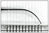
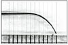
To assess instrumentation of curved canals, clear resin blocks were used in this study. These blocks were chosen because of the standardized shape, size, taper, and curvature of all roots15,16). Weine et al.2) have validated the credibility of a resin block as an ideal experimental model for quantitative and qualitative analyses of endodontic preparations. Used for the study of rotary instruments, however, the resin material is not ideal because it does not cut in the same way as dentin. Many rotary instruments do not have sharp cutting edges, but remove dentin by a grinding action. The effect of this grinding action in resin is unknown, but heat generated may sometimes soften the resin material. Nevertheless for a study like this it is the only material presently available, and it is very useful if the results are properly interpreted17).
The ProTaper series is comprised of 3 "shaping" and 3 "finishing" instruments. A unique feature of the shaping files is their progressively taper design, which clinically serves to significantly improve flexibility and cutting efficiency; it typically reduces the number of recapitulations needed to achieve length, especially in tight or more curved canals. Additionally, a progressively tapered file engages a smaller zone of dentin, which reduces torsional loads, file fatigue, and the potential for breakage18).
Manufacturers of the ProTaper recommended that finishing files are not suitable for novice operators, because the nature of finishing file is too aggressive to create various defects. Therefore we used ProFile systems as finishing files of ProTaper after using of shaping files of ProTaper. It was the Hybrid technique that used in this study.
Mandel et al.10) found that there was a learning period in novice operators and they were not novice operators any more after they got through a learning period. Only three root canals were instrumented by one novice operator in this study.
Adobe Photoshop provides a convenient means of comparing pre-instrumentation images and post-instrumentation images. Using the Adobe Photoshop, photographs of pre-instrumentation and post-instrumentation could be superimposed. Changing the opacity of the layers allowed visualization of the movement of the instrumented canal at each level19).
This experiment used a centering ratio formula to evaluate canal transportation. According to this formula, centering ratio approaches zero as X1 and X2 become closure. Zero is an indication of perfect canal centering and no canal transportation13). All groups produced some canal transportation in this study.
Weine et. al.2) demonstrate formation of hourglass-shaped preparations in curved canals. It is a reflection of the tendency for instruments to straighten. It was expected that transportation towards the inner curvature would occur in the middle segment and the outer curvature in the apical segment13).
From this study, it was found that Ni-Ti rotary files caused taper widening of the canal space to the inner portion of the curvature at 4, 5, 6, 7 mm level and to the outer portion of the curvature at 1, 2, 3 mm level. At 3 mm level in novice operators with ProTaper and at 3 mm level in endodontists with Hybrid, the opposite result was obtained. These results are similar to other reports with Ni-Ti rotary files20,21). Bryant et al.20) identified 'the outer widening' in their study using ProFile .04 and .06 taper instruments. Since the .06 instrument is stiffer than the .04 file, it has a tendency to straighten the curved canal. The inner widening of middle level of the canal and the outer widening of the apical canal can be the result of this phenomenon. To prevent this phenomenon, Griffiths et al.21) recommended that clinicians need to restrict the use of larger and stiffer instruments in the deeper portion of canal.
The centering ratio showed that there was significant differences between operators at 3 mm level in all groups. However, the values of centering ratio were closer to zero than those at other levels from the apex therefore it did not induce severe canal transportation. At the level of apex, apical transportations in novice operators with ProTaper showed greater value than those in endodontists. This result done by novice operators with ProTaper reflects that canal shaping at the apical area is difficult, because of great cutting efficiency of ProTaper.
Time efficiency is one aspect of the shaping efficiency. The instrumentation time may be influenced by the canal shape, the type of instruments used, the number instruments changed, the amount of irrigation and recapitulation, apical preparation size, and the skill of the individual operator. Novice operators spent significantly more time than endodontists and canal preparation time in profile technique was significantly longer than in the other techniques.
Therefore hybrid use of ProFile and ProTaper is recommended to novice operators than more time consuming Profile technique.
In this study, instrumentation time of ProFile was longer than those in other study. Because resin block used in this study had severe curvature (41.4 degree by Schneider's method), we shaped the canal with care. This result is similar to other report with same resin block17).
Kavanagh and Lumley22) and Bryant et al.20) found that the use of Ni-Ti rotary instruments was effective and produced good canal shapes. Other studies23,24) partly supported their observations. Gluskin et al.25) found that novice dental students were able to prepare curved root canals with Ni-Ti rotary files with less transportation and greater conservation of tooth structure, compared to canals prepared with hand instruments. Therefore novice operators can prepare canals with Ni-Ti rotary files in safe and efficient way if educated properly.
Further research is needed to evaluate the efficiency of Ni-Ti files in human teeth shaped by novice operators.


 XML Download